The human eye: how vision works

In a day, our eyes silently absorb myriad visual experiences, from the soft glow of a bedside lamp to the vibrant hues of a sunrise, showcasing their remarkable ability to convert light rays into the visual symphony that colors our daily lives.

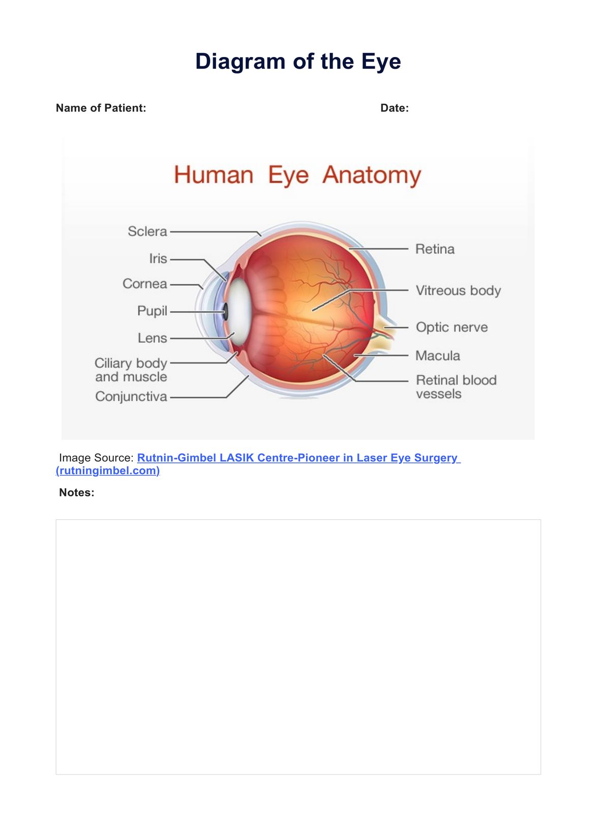
Light rays bounce off the object we are looking at. Then, the journey begins when light passes through the cornea, a transparent outer layer acting as a natural lens that focuses light to make things look sharp. Next, it passes through the pupil, the dark circle in the eye.

The iris, the colored part of the eye, adjusts like a camera aperture, controlling incoming light. Our eyes naturally adapt to different light conditions. The iris controls the widening or narrowing of the pupil, ensuring the right amount of light enters. In low light, the pupil widens for acute vision, while in bright light, it narrows to prevent surplus light. Behind is the lens, a vital part of the eye's anatomy, that refracts incoming light onto the light-sensitive layer — the retina (National Eye Institute, 2022).

The retina is home to two types of cells: rod cells, specialized for dim light and peripheral vision, and cone cells, responsible for central vision and distinguishing colors and essential for receiving a sharp, accurate image. These cells convert light into electrical impulses, a crucial step in processing all the visual information (Moorfields Eye Hospital NHS Foundation Trust, 2022).

Nerve fibers carry these electrical impulses to the optic disc, a transparent structure at the back of the eye. It marks where nerve fibers converge to form the optic nerve, the gateway through which all visual information embarks on its journey to the brain. The optical disc identifies the same way for every human eye, playing a pivotal role in our eye anatomy.

Challenges may arise in this intricate process of light and perception. An operation becomes necessary when a cloudy lens affects vision clarity. This cataract operation involves replacing the clouded lens with an artificial plastic lens, restoring clear vision.

In the silent storytelling of our eyes, specialized cells translate light into electrical energy. This energy, transformed into visual messages, creates our vision. From the nuances of color to the contrasts defining our world, our eyes, with their transparent structure and precise vision capabilities, play an integral role in shaping our perception.

Printable Diagram of the Eye

Download this Diagram of the Eye that illustrates the practical application of an eye test chart in real-life situations.

When is it useful to have an Eye Diagram?

Understanding the intricate workings of the human eye is vital for maintaining optimal vision and addressing potential issues. An eye diagram serves as a valuable tool to comprehend the complexities of this essential sensory organ. It becomes beneficial in various scenarios, offering insights into eye anatomy, visual processes, and potential concerns.

Visual Education and Patient Understanding

Eye diagrams provide a clear visual representation of the complex structures within the eye. An eye diagram is an invaluable educational resource for individuals seeking to understand the role of light-sensitive cells, optic nerves, blood vessels, and the transparent structures that refract the light entering the retina. It aids comprehension and empowers individuals to participate actively in their eye health.

Diagnostic and medical communication

1. Assessing and communicating eye conditions

In a medical context, eye diagrams are indispensable for clinicians communicating with patients about eye conditions. When explaining issues like cataracts or the impact of how much light enters the eye, a visual aid simplifies complex concepts. It bridges medical terminology and patient understanding, ensuring individuals comprehend the factors affecting their vision.

2. Guiding surgical procedures

During a cataract operation, for instance, surgeons utilize diagrams to illustrate the procedure involving the replacement of the natural lens. The diagram helps convey the process of implanting an artificial lens, offering patients a tangible understanding of the surgery and the role of different primary colors in their visual perception.

Vision care and optometric practice

1. Promoting eye health

Eye diagrams are instrumental in promoting eye health and preventive care. By visualizing how light-sensitive structures in the eye absorb excess light or how the primary colors contribute to vision, individuals gain insights into practices that support healthy eyes. This includes understanding how different primary colors impact the sharpness of images on the retina.

2. Educating on eye adaptation

For those exploring the effects of light on vision, an eye diagram facilitates learning about the iris's role in adjusting to different light conditions. It aids in comprehending how the eye's middle layer adapts to the amount of light, ensuring precise vision in varying environments.

In summary, an eye diagram proves helpful in diverse scenarios—educational settings for visual literacy, medical contexts for diagnostic clarity, and vision care for promoting eye health awareness. Its value lies in making the complexities of the human eye accessible, fostering understanding, and empowering individuals to actively engage in maintaining their vision.

Eye Diagram example (sample)

The provided Eye Diagram Example, accessible for download and examination, goes beyond a simple outline. It illustrates the practical application of an eye test chart in real-life situations. Secure your copy today by previewing the sample or selecting the "Download Example PDF" button.

Download this Diagram of the Eye example:

Eye Diagram example (sample)

Signs and symptoms someone needs an eye test

Regular eye tests are crucial for maintaining optimal vision and overall eye health. While the absence of apparent issues may suggest good vision, signs, and symptoms can indicate the need for an eye test. Paying attention to these cues allows individuals to address potential vision issues promptly.

  1. Vision changes: If you notice sudden changes in your vision, such as blurred or distorted sight, difficulty focusing, or persistent double vision, it's a clear signal to schedule an eye test.
  2. Eye strain and discomfort: Frequent headaches, eye strain, or discomfort can indicate an underlying vision problem. Prolonged screen time, reading in poor lighting, or uncorrected vision issues may contribute to these symptoms.
  3. Light sensitivity and glare: Increased sensitivity to light or experiencing excessive glare, especially during activities like driving at night, may suggest issues such as cataracts or other eye conditions.
  4. Difficulty adjusting to darkness: Struggling to adapt to low-light conditions or experiencing night vision difficulties may indicate an underlying issue. Conditions like nyctalopia, commonly known as night blindness, can be identified through an eye test, ensuring appropriate measures are taken to address the challenges associated with dim lighting.
  5. Persistent eye redness or irritation: Persistent redness, itchiness, or irritation in the eyes may indicate various eye conditions, including allergies, dry eye syndrome, or infections.
  6. Family history of eye conditions: Regular eye tests, especially for those with a family history, can help detect and manage potential concerns early, preserving vision and preventing complications.

Other helpful resources

Delve into the intricacies of the human eye with Carepatron's resources. Explore our Diagram of the Eye & Example for clear vision and efficient visual messages. Discover more resources below."

  1. Eye Test Chart Template: Evaluate your vision with our precise eye test chart template, designed to assess the thin transparent capsule, distinguish colors, and ensure clear vision.
  2. Ophthalmologic Exam Template: Conduct a comprehensive ophthalmologic exam using our template, which examines the same way the human eye processes visual information, ensuring a compassionate evaluation of eye anatomy.
  3. Vision Screening Template: Utilize our vision screening template to focus on the human retina, distinguish colors, and evaluate eye anatomy, all geared towards maintaining clear vision.
  4. Amsler Grid Chart Template: Assess your vision with the Amsler grid chart, a valuable tool for the human eye, maintaining a compassionate approach to distinguishing colors and ensuring precise vision.

References

Moorfields Eye Hospital NHS Foundation Trust. (2022, August 18). Anatomy of the eye. https://www.moorfields.nhs.uk/content/anatomy-eye

National Eye Institute. (2022, June 9). The Visual System: How Your Eyes Work [Video]. YouTube. https://www.youtube.com/watch?v=TY1giZgddAs

How does the Diagram of the Eye relate to the human body?

Commonly asked questions

How does the Diagram of the Eye relate to the human body?

The diagram illustrates the intricate structures within the eye, showcasing its role as a vital organ in the human body.
What are the benefits of using the anatomy of the eye?

Healthcare professionals, such as optometrists and ophthalmologists, can utilize the diagram to communicate visually with patients about eye conditions. It aids in simplifying complex concepts, facilitating better patient understanding, and fostering a collaborative approach to eye care.

Who typically uses the Diagram of the Eye, and for what purpose?

Eye care professionals, educators, and students commonly use this diagram to understand, teach, and learn about the complexities of the eye's anatomy and its functions.

Join 10,000+ teams using Carepatron to be more productive

One app for all your healthcare work