Ulnar nerve pathway
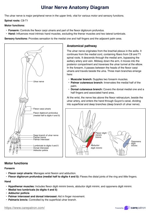
The ulnar nerve, a major peripheral nerve of the upper limb, is crucial in motor and sensory functions. Originating from the C8 and T1 spinal nerve roots, it is a continuation of the medial cord of the brachial plexus. This nerve provides functionality and sensation to muscles in the forearm and hand.
The ulnar nerve originates from the axilla (armpit) region as it exits the brachial plexus, running down the medial aspect of the upper arm alongside the brachial artery. At the midpoint of the arm, the ulnar nerve descends through the medial intermuscular septum, entering the posterior compartment. This pathway continues as the nerve passes posteriorly to the medial head, traversing through the cubital tunnel—a space between the medial epicondyle of the humerus and the olecranon of the ulna, near the elbow joint.
In the forearm, the ulnar nerve pierces the two heads of the flexor carpi ulnaris muscle and travels deep along the ulna, where three significant branches arise:
- Muscular branch: Innervates the flexor carpi ulnaris and the medial half of the flexor digitorum profundus.
- Palmar cutaneous branch: Provides sensation to the medial half of the palm.
- Dorsal cutaneous nerve branch: Supplies the dorsal surface of the medial one and a half fingers and associated hand area.
At the wrist, the ulnar nerve lies superficial to the flexor retinaculum (also known as the transverse carpal ligament), adjacent to the ulnar artery. It enters the hand through Guyon's canal, where it divides into superficial and deep branches.
The ulnar nerve superficial branch innervates the palmaris brevis muscle and provides sensation to the medial one and a half fingers. The deep branch of the ulnar nerve supplies most of the intrinsic muscles of the hand, including the hypothenar muscles, the medial two lumbricals, the adductor pollicis, the flexor pollicis brevis, and the interossei muscles (both palmar and dorsal).


