What is the Spurling's Test?
The Spurling test, initially named Spurling's neck compression test by neurosurgeons Roy Glen Spurling and William Beecher Scoville, was introduced in 1944 to evaluate "radiculitis." It is also known as the foraminal compression test, Spurling's neck compression test, or the quadrant test.
Spurling's Test is mainly used to detect nerve root compression in the cervical spine (Shelow et al., 2020). Numerous disorders, including ruptured discs, bone spurs, and degenerative disc degeneration, might contribute to this compression. Head and neck pain, arm pain, numbness, tingling, and paralysis can result from cervical spine compression.
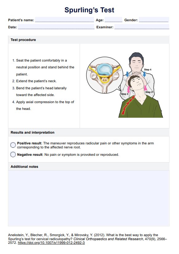
The original procedure for Spurling's Test was developed in 1944 and is no longer considered the proper technique (Jones & Miller, 2020). According to a study by Anekstein et al. (2012), the procedure that can provoke the most symptoms related to unilateral cervical radiculopathy involves the following steps:
- With the patient in a seated position, extend the neck.
- Position the patient's head at lateral flexion towards the affected side.
- Apply downward pressure.
This test has a low sensitivity at 0.50, but it has a high specificity at 0.88 (Wainner et al., 2003).


