How does our Median Nerve Anatomy Diagram work?
To help you master the median nerve anatomy, we created a Median Nerve Diagram Template you can download by clicking the button and using the links in this guide.
Follow our provided template by following our step-by-step instructions below on how to use our diagram template:
Step 1: Have a basic understanding
Start with a foundational understanding of the median nerve’s origin and course from the lateral and medial cords of the brachial plexus. Understanding the nerve roots and their contributions is essential for grasping the entire nerve structure.
Step 2: Study the anatomical course
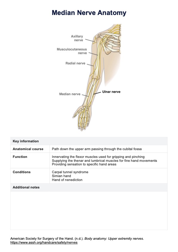
Study the anatomical course of the median nerve, which is a path down the upper arm at the elbow joint, passing through the cubital fossa alongside the ulnar nerve and continuing into the forearm and hand.
Step 3: Understand the functional anatomy
Understand the muscles innervated by the median nerve, which are most forearm flexor muscles, thenar muscles, and lumbrical muscles. Also, it is important to recognize the role of the anterior interosseous nerve, which is connected to the median nerve, in controlling some of these muscles.
Finally, learn the sensory distribution areas of the median nerve, which include the palmar side of the thumb, index, middle, and part of the ring finger. The palmar cutaneous branch of the median nerve is crucial for this sensory innervation.
Step 4: Check clinical relevance
Familiarize yourself with common clinical conditions involving the median nerve and their clinical signs and symptoms, such as sensory deficits and motor dysfunctions affecting the index and middle fingers, thumb opposition, and grip strength. Understand how median nerve injury can lead to these conditions.