Abdominal Examination

Download our comprehensive Abdominal Examination PDF guide for detailed procedures and reporting, ideal for healthcare professionals and students.

By Emma Hainsworth on May 13, 2024.

Fact Checked by Ericka Pingol.

Use Template

What is an abdominal examination?

An abdominal examination is a clinical procedure performed by a healthcare provider to assess the organs and structures of the abdominal region. This examination is an essential part of the physical assessment of a patient, as it can help diagnose a wide range of conditions that may affect the digestive system, the urinary system, and other organs located in the abdomen. The examination typically includes several key components:

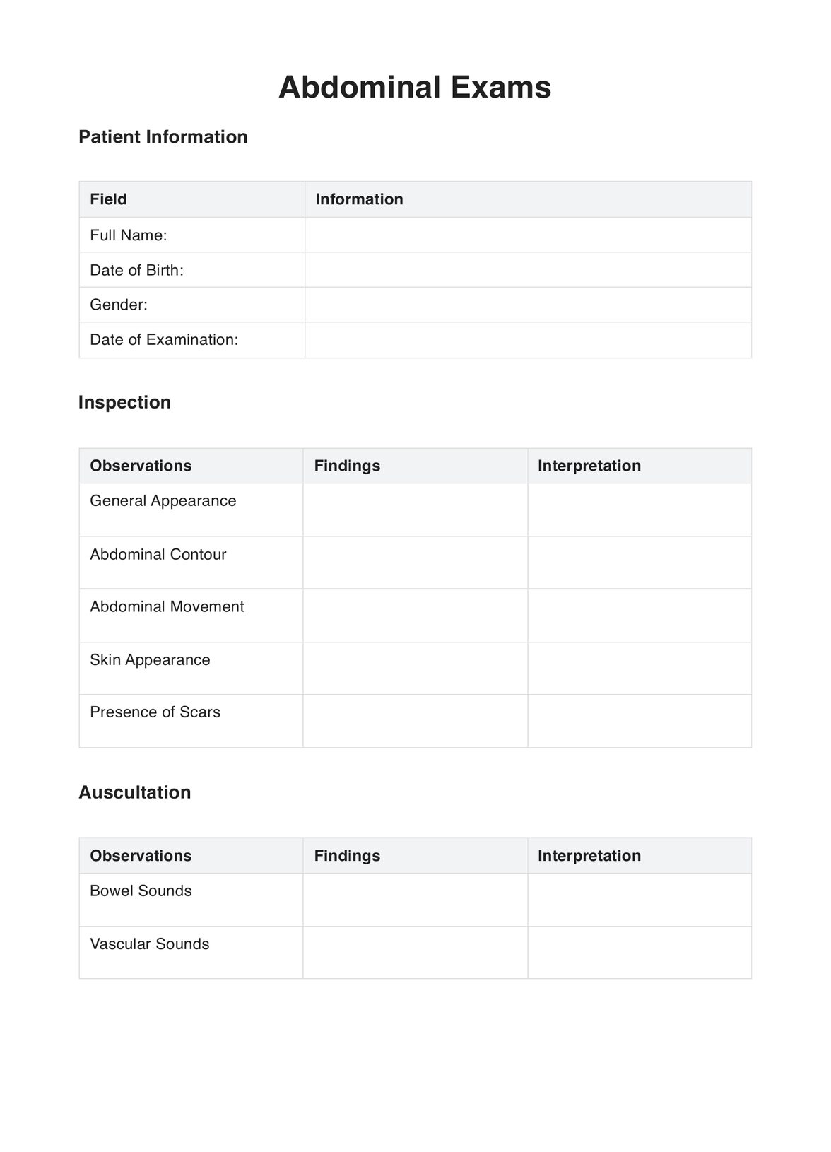
  • Inspection: The examiner looks at the abdomen to identify any visible signs such as swelling, scars, skin changes, abnormal movements (like visible peristalsis), or the shape and contour of the abdomen.
  • Auscultation: Using a stethoscope, the examiner listens to the sounds within the abdomen, particularly bowel sounds. This can give clues about the functioning of the intestines and the presence of any abnormal activity.
  • Percussion: Tapping on the abdomen helps to determine the size of the organs, detect the presence of fluid or gas, and assess the tone of the abdominal wall.
  • Palpation: The examiner gently feels the abdomen with their hands to check for tenderness, swelling, and the size and shape of the abdominal organs. This part of the examination can provide information about the liver, spleen, kidneys, and other structures.
  • Special tests: Depending on the findings and the patient's symptoms, the examiner might perform additional tests like checking for rebound tenderness (which can indicate inflammation), testing for fluid in the abdominal cavity (ascites), or assessing for specific syndromes like Murphy's sign (related to the gallbladder).

An abdominal examination aims to gather information about the health of the patient's abdominal organs, including the stomach, liver, gallbladder, spleen, kidneys, intestines, and other structures. It helps diagnose infections, tumors, inflammations, fluid accumulation, and organ enlargement. The examination is performed systematically to ensure that no aspect of the abdomen is overlooked and is usually conducted while the patient is lying down.

Printable Abdominal Examination

Download this Abdominal Examination to assess the of the abdominal region.

Clinical signs your patients need an Abdominal Exam

Several vital signs and symptoms may indicate the need for an abdominal examination when evaluating patients in a clinical setting. This examination is crucial for assessing the abdominal organs and structures, including the abdominal muscles, abdominal wall, and the contents of the abdominal cavity. Here are some clinical signs to look out for:

  • Abdominal pain: One of the most common indications for an abdominal exam is abdominal pain. This can range from acute abdominal pain, often requiring immediate attention, to chronic pain, which may indicate ongoing issues such as chronic liver disease.
  • Abdominal distension: Distension or swelling of the abdomen could suggest various underlying pathologies, from fluid accumulation (ascites) to bowel obstruction. It's essential to evaluate the entire abdomen, including all abdominal quadrants.
  • Changes in bowel sounds: Both hyperactive and absent bowel sounds can be significant. Hyperactive sounds might indicate bowel obstruction or inflammatory bowel disease, while absent bowel sounds could suggest peritoneal inflammation or severe liver disease.
  • Visible changes in the abdominal wall include changes in the abdominal muscles, such as bulging due to an umbilical hernia or visible scars from previous abdominal surgery.
  • Presence of bruits: Auscultation for bruits, especially over the renal arteries, can indicate vascular issues like renal artery stenosis. Bruits over the abdominal aorta might suggest turbulent blood flow, often associated with an abdominal aortic aneurysm.
  • Rebound tenderness: This is a clinical sign of peritoneal irritation, often found in acute abdomen conditions like appendicitis.
  • Palpable masses: During abdominal palpation, any abnormal masses in the abdomen, such as an enlarged liver (hepatomegaly) or spleen (splenomegaly), can be indicative of underlying pathology.
  • Jaundice or other skin changes: These could be signs of liver disease, which would warrant a detailed examination of the liver, including palpation for a palpable liver edge.
  • Urinary or renal complaints: Symptoms such as flank pain or changes in urination can necessitate a thorough examination of the kidneys, including checking for enlarged or cystic kidneys.
  • Gastrointestinal symptoms: Chronic symptoms like diarrhea, constipation, or blood in the stool may require a comprehensive abdominal exam, including a digital rectal examination.
  • History of portal hypertension or alcohol abuse: Such a history may prompt a detailed liver examination due to the risk of severe liver disease.
  • Routine physical examinations: Even without specific symptoms, routine physical examinations often include an abdominal exam to check for unnoticed issues.

Abdominal Exam components

Palpation for lymphadenopathy

  • Purpose: To check for enlarged lymph nodes, which can indicate infection, inflammation, or malignancy.
  • Method: Gentle palpation in areas with lymph nodes in the abdominal region.

Palpating the liver

  • Purpose: To assess liver size, texture, and presence of tenderness.
  • Method: Palpation is usually done starting in the right lower quadrant (RLQ) and moving toward the liver area in the upper quadrant.

Palpating the gallbladder

  • Purpose: To detect gallbladder enlargement or tenderness indicative of conditions like cholecystitis.
  • Method: Palpation is performed in the upper right quadrant, often using Murphy's sign.

Palpating the spleen

  • Purpose: To assess for splenomegaly, which can indicate various underlying conditions.
  • Method: Palpation starts in the RLQ, moving towards the left upper quadrant (LUQ).

Palpating the bladder

  • Purpose: To check for bladder enlargement or tenderness.
  • Method: Palpation in the lower abdomen, particularly when the bladder is expected to be complete.

Percussing the liver

  • Purpose: To estimate liver size and detect abnormalities in liver density.
  • Method: Percussion starts in the RLQ, moving towards the right upper quadrant.

Percussing the spleen

  • Purpose: To check for splenomegaly.
  • Method: Percussion of Traube's space (left lower chest area) for changes in sound from tympany to dullness.

Percussing the bladder

  • Purpose: To determine bladder size and presence of urine.
  • Method: Percussion is done in the lower abdomen, particularly over the bladder region.

Assessing bowel sounds

  • Purpose: To evaluate intestinal motility.
  • Method: Auscultation is performed using a stethoscope over various quadrants of the abdomen to listen to bowel sounds.

Each of these steps is integral to a thorough abdominal examination, providing valuable information about the health of the abdominal organs and identifying potential abnormalities.

How does an Abdominal Examination work?

The abdominal examination is a clinical process that involves several steps to assess the health of the abdominal organs and to identify any abnormalities. Here's a brief overview of how it works:

  1. Preparation: The patient is usually asked to lie down in a supine position with their abdomen exposed. This helps in relaxing the abdominal muscles, providing better access for examination.
  2. Inspection: The examiner visually inspects the abdomen for any signs of swelling, scars, skin changes, visible movements (like peristalsis), and the overall contour and symmetry.
  3. Auscultation: Before any palpation or percussion, which might alter bowel activity, the examiner listens with a stethoscope to the bowel sounds in various quadrants of the abdomen. This step assesses the intestinal activity and can detect abnormalities like bowel obstruction.
  4. Percussion: This involves tapping on the abdomen to assess the size of the organs like the liver and spleen and to detect the presence of fluid or gas. Different sounds can indicate various conditions; for instance, a dull sound might suggest fluid accumulation (ascites).
  5. Palpation: The examiner gently presses the abdomen to feel for any masses, tenderness, or organ enlargement. This step is crucial for assessing structures like the liver, spleen, kidneys, and bladder. Palpation is usually done systematically, starting from areas furthest from the pain.
  6. Special maneuvers: Depending on initial findings, tests like checking for rebound tenderness (a sign of peritonitis) or performing Murphy's sign (for gallbladder disease) might be included.
  7. Documentation and report: The findings from each step are carefully documented, often using a standardized format like a PDF. This record includes observations from all examination steps and helps diagnose, plan treatment, and monitor progress.

Overall, an abdominal examination is a fundamental component of a physical examination, providing vital information about the health of the abdominal organs and guiding further medical investigation or treatment.

Abdomen Examination example (sample)

For those looking to understand how an abdomen examination is conducted, you can now access a practical example. We offer a free abdomen examination sample that is available for download. This example is an excellent resource for medical professionals, students, or anyone interested in learning about the intricacies of performing an abdominal assessment.

Download this free Abdomen Examination example here:

Abdomen Examination example (sample)

Why use Carepatron as your Abdomen Examination software?

Using Carepatron as your abdomen examination software offers several advantages, particularly in enhancing the efficiency and accuracy of abdominal assessments. Here's why it's an excellent choice for healthcare providers:

  • Comprehensive clinical documentation: Carepatron provides detailed clinical documentation capabilities, allowing healthcare professionals to record, update, and track all relevant patient information and examination findings related to abdominal health. This feature is crucial for accurate diagnosis and treatment planning.
  • Integrated electronic health records (EHR): With its EHR system, Carepatron ensures that all patient data, including abdomen examination results, medical history, and treatment plans, are securely stored and easily accessible. This integration is vital for maintaining continuity of care and effective patient management.
  • Efficient appointment scheduling: Carepatron's appointment scheduling feature streamlines arranging follow-up visits, which is essential for ongoing abdomen health monitoring and management.
  • Telehealth capabilities: Offering telehealth services can benefit patients who need regular abdomen health check-ups but cannot visit the clinic frequently. This feature enhances patient access to care and allows for remote monitoring.
  • Customizable forms and templates: Carepatron allows healthcare providers to customize forms and templates to specific needs, which is particularly useful for tailoring documentation to the unique requirements of abdomen examinations.

By choosing Carepatron for abdomen examination needs, healthcare providers benefit from a comprehensive, secure, and user-friendly platform that enhances patient care and improves operational efficiency.

Practice Management Software
What is the significance of assessing abdominal wall muscles during a physical examination?

Commonly asked questions

What is the significance of assessing abdominal wall muscles during a physical examination?

During a physical examination, assessing the abdominal wall muscles is crucial for detecting abnormalities in the patient's abdomen. This includes examining the strength and tone of the abdominal musculature. The evaluation helps identify issues like muscle weakness, hernias, or abdominal pathology. It often involves techniques like palpation and observing the patient's abdominal muscles while they take a deep breath or perform specific movements.

How are deep palpation and light palpation used to examine abdominal organs?

Deep and light palpation are critical techniques in clinical medicine for examining abdominal organs. Light palpation assesses surface-level characteristics like tenderness. In contrast, deep palpation allows for a more detailed physical examination of intra-abdominal organs such as the liver, spleen, and kidneys. For instance, deep palpation can help detect an abdominal mass or the enlarged liver edge in liver disease, while light palpation helps assess superficial tenderness or muscular resistance.

What does the absence of bowel sounds indicate in abdominal auscultation?

Absent bowel sounds during abdominal auscultation can be a significant clinical finding. Usually, bowel sounds are present due to the movement of the intestines. Absent or significantly reduced bowel sounds may indicate a lack of intestinal activity, which can be associated with conditions like severe liver disease, bowel obstruction, or peritonitis. In contrast, hyperactive bowel sounds might suggest diarrhea or early bowel obstruction.

Join 10,000+ teams using Carepatron to be more productive

One app for all your healthcare work