Heart Valve Test

Learn more about the different types and symptoms of heart valve tests, their benefits, and how they help diagnose heart valve problems with this comprehensive guide.

By Joshua Napilay on May 15, 2024.

Fact Checked by Ericka Pingol.

Use Template

How do the heart valves function?

The heart valves play a vital role in the function of the heart. The heart has four valves: the mitral, pulmonary, tricuspid, and aortic. All four valves open and close to help move blood from one area to another.

Two of the valves, the mitral and the tricuspid valves, move blood from the heart's upper chambers (the atria) to the lower chambers (the ventricles). The other two valves, the aortic and pulmonary valves, move blood to the lungs and the rest of the body through the ventricles.

The job of the valves is to move the blood through the heart. When the two atrium chambers contract, the tricuspid and mitral valves open, allowing blood to move to the ventricles. When the two ventricle chambers contract, they force the tricuspid and mitral valves to close as the pulmonary and aortic valves open.

The valves should prevent the blood that leaves the ventricles from traveling to the body from flowing back to the left ventricle into the atria. The cusps help keep blood flowing, and the valves create a tight seal between blood vessels, which allows blood to flow from the left ventricle in the correct direction. When the heart valves open and close, they make sounds we know as our heartbeat.

Printable Heart Valve Test Template

Download this Heart Valve Test Template to streamline and organize essential data for comprehensive assessments of heart valve function, facilitating efficient analysis and informed decision-making in cardiovascular care.

What is an echocardiogram?

An echocardiogram, also known as an echocardiograph or diagnostic cardiac ultrasound, is a test that uses high-frequency sound waves (ultrasound) to create pictures of the heart. This noninvasive test allows healthcare providers to assess the heart's structure, function, and blood flow through the valves. There are several types of echocardiograms, including:

  • Transthoracic echocardiography
  • Stress echocardiography
  • Transesophageal echocardiography
  • Three-dimensional (3D) echocardiography

The test is performed to diagnose various heart conditions, including valve disease, cardiomyopathy, and other heart-related issues. Echocardiography is particularly useful for evaluating blood flow across the heart's valves and detecting problems such as regurgitation, stenosis, or abnormal growths around the valves. The test is painless and has no side effects, making it a valuable tool for diagnosing and monitoring heart health.

Heart Valve Test example (sample)

This downloadable template is a comprehensive guide for medical professionals requesting cardiac imaging assessments for patients with potential heart valve abnormalities. Feel free to customize the template according to your specific clinic or hospital requirements, ensuring that it aligns with the unique needs of your medical practice.

Download this Heart Valve Test Template example:

Heart Valve Test example (sample)

How is heart valve disease diagnosed?

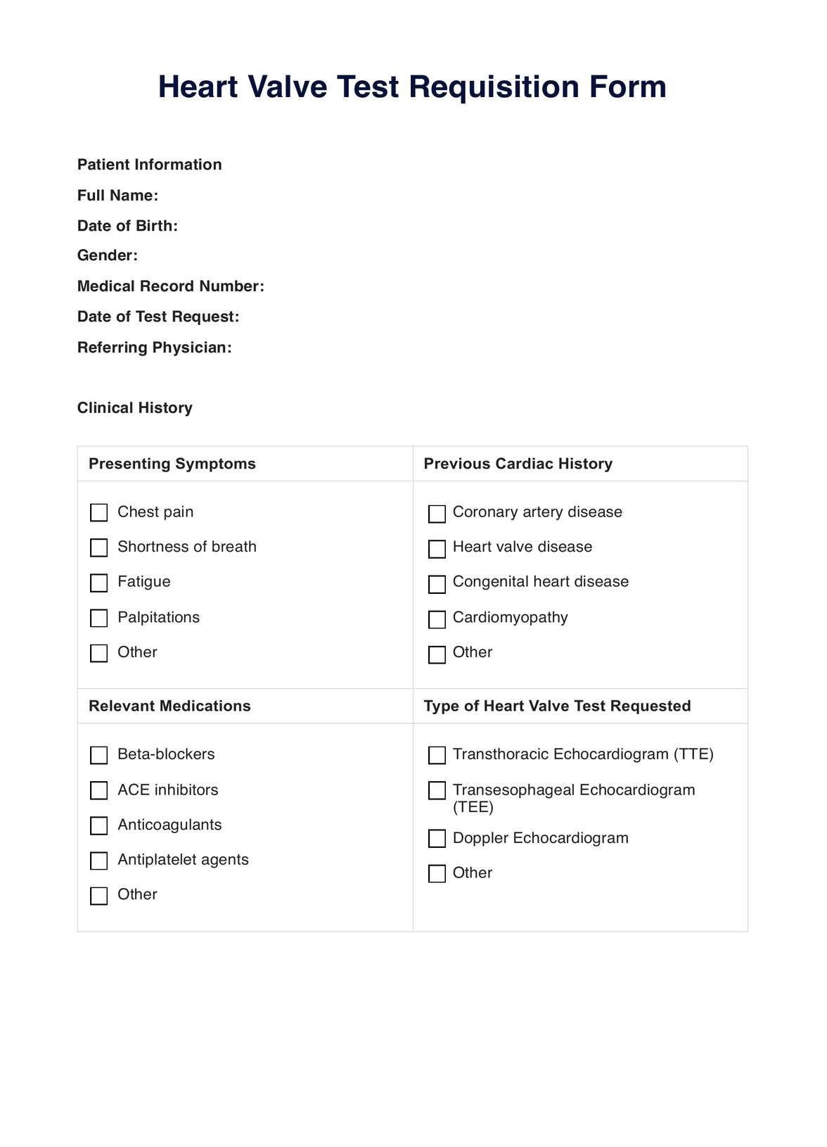
Heart valve disease symptoms can be diagnosed using a combination of physical examination, medical history, and various tests. The diagnostic process typically includes the following steps:

Physical examination

During a physical examination, a healthcare professional listens to the patient's heart with a stethoscope to detect any unusual sounds or murmurs, which can indicate valve problems. This is the first diagnostic measure for heart valve problems and is a valuable tool for screening and diagnosis. Other diagnostic tests, such as echocardiography, may be used to evaluate heart valve function further and diagnose heart valve diseases and problems.

Medical history

Medical history is essential to assessing the likelihood of heart valve disease. The patient's symptoms, risk factors, and health history are considered to determine if they are at risk for heart valve diseases or problems.

Electrocardiogram (ECG or EKG)

An electrocardiogram (ECG or EKG) is a test that records the heart's electrical activity and can help identify any abnormalities related to valve problems.

The test uses electrodes placed on the patient's chest and limbs, which detect electrical signals generated by the heart. ECGs are noninvasive, painless, and can provide information about the heart's electrical activity, such as heart rate, rhythm, and potential abnormalities.

Echocardiogram (Echo)

An echocardiogram (echo) is a noninvasive test that uses high-frequency sound waves (ultrasound) to create images of the heart. It provides information about the heart's structure, function, and blood flow through the valves.

There are several types of echocardiograms, including transthoracic, transesophageal, stress, and three-dimensional (3D) echocardiography. The test is commonly used to diagnose heart conditions, including cardiomyopathy and valve disease.

Two-dimensional echocardiogram (2D Echo)

A two-dimensional echocardiogram (2D Echo) is an advanced form of echocardiography that provides more detailed images of the heart's structure and function. It assesses the heart's overall health, evaluates abnormal heart structures, and detects various heart conditions, including valve problems. The test uses a transducer that sends high-frequency sound waves to the heart, displaying the resulting images on a monitor.

Exercise test

An exercise test, also known as a stress test, assesses the heart's response to physical activity and detects any changes in valve function. This test involves walking on a treadmill or riding a stationary bike while checking the heart.

Chest X-rays, CT scans, and cardiac catheterization

Chest X-rays, CT scans, and cardiac catheterization are additional tests that may be used to evaluate the heart valves and the patient's overall cardiac health further.

Cardiac catheterization is an invasive procedure to evaluate heart function, while CT scans and chest X-rays provide detailed images of the heart and surrounding structures. These tests can help diagnose and assess heart valve problems, providing valuable information for treatment planning and management.

What causes heart valve damage?

Heart valve damage can be caused by several factors, including:

  • Aging: Changes in the heart valve structure due to aging can lead to valve damage.
  • Coronary artery disease and heart attack: Narrowing the coronary arteries can reduce blood flow to the heart, causing damage to the heart valves.
  • Heart valve infection: Infections like rheumatic fever or blood infections can cause heart valve damage.
  • Congenital disabilities: Some people are born with heart valve disease, a condition called congenital heart valve disease.
  • Infections: Certain infections, such as rheumatic fever or blood infections, can damage the heart valve.
  • High blood pressure: Excessive pressure on the heart valves can cause damage over time.
  • Mechanical or artificial heart valves: Prior treatment for heart valve problems, such as surgery to repair or replace a valve, can cause damage to the heart valve.

Other diagnostic tests and procedures

Here are some additional diagnostic tests and procedures:

  • Exercise stress test: Measures the heart's response to physical exertion, helping to diagnose coronary artery disease and assess overall cardiac function.
  • Nuclear stress test: Combines stress testing with injecting a small amount of radioactive substance to create images of blood flow to the heart.
  • Holter monitor: A portable device worn by the patient for 24 to 48 hours, continuously recording the heart's electrical activity to detect irregularities.
  • Event recorder: Similar to a Holter monitor but used for extended periods. The patient activates the device when experiencing symptoms, allowing for intermittent monitoring over weeks or months.
  • Cardiac catheterization (angiography): An invasive procedure involves the insertion of a catheter into the coronary arteries to assess blood flow, measure pressures, and identify blockages or narrowing.
  • Coronary CT angiography (CTA): Noninvasive imaging technique that uses computed tomography to visualize the coronary arteries and assess for blockages or abnormalities.
  • Cardiac MRI (magnetic resonance imaging): Provides detailed images of the heart's structure and function, aiding in diagnosing various cardiac conditions, including heart valve disorders, cardiomyopathies, and congenital heart diseases.
  • Cardiac biomarkers: Blood tests measure substances released into the blood when the heart is damaged. Examples include troponin and creatine kinase-MB (CK-MB).
  • Lipid profile: Assesses cholesterol levels in the blood, providing information about cardiovascular risk.
  • Coronary calcium scoring: CT scans to assess the amount of calcium in the coronary arteries, helping to estimate the risk of coronary artery disease.
  • Ambulatory blood pressure monitoring: 24-hour monitoring comprehensively assesses blood pressure patterns daily and night.
  • Tilt table test: Evaluates the autonomic nervous system by monitoring heart rate and blood pressure changes in response to changes in body position.

Electrophysiological studies (EPS): Invasive procedures to study the heart's electrical system, particularly the conduction of electrical impulses through the heart.

The importance of medical histories and physical exams

Medical histories and physical exams are foundational components of healthcare that play a crucial role in the diagnostic process, treatment planning, and overall patient care. They are fundamental tools for healthcare professionals to gather comprehensive information about a patient's health, identify potential risk factors, and establish a basis for ongoing medical management.

Here are some key aspects highlighting the importance of medical histories and physical exams:

  • Early detection and prevention
  • Diagnostic clues
  • Treatment planning
  • Communication and trust
  • Holistic approach
  • Screening for red flags
  • Monitoring changes over time
  • Cost-effective healthcare

Why is an exercise test sometimes needed to assess a valve problem?

An exercise test, also known as a stress test, is sometimes needed to assess a valve problem for several reasons:

  • Evaluate functional disability: Exercise testing objectively assesses functional disability, which is crucial in patients with valvular heart disease.
  • Assess symptoms: Exercise testing can help identify the valve and ventricle dynamics and evaluate changes in forward output, retrograde flow, and pulmonary pressures.
  • Detect hidden symptoms: Some patients with valvular heart disease may not realize they have limited activity over time or attribute changes to "normal aging." Exercise testing can help detect these symptoms.
  • Evaluate heart response: Exercise testing can help evaluate changes in blood pressure and symptoms and the heart's response to a more challenging workload.
  • Determine disease severity: Exercise tests can show how the heart responds to physical activity and whether valve disease symptoms occur during exercise, helping to determine the severity of heart valve disease.

How long does it take to recover from heart valve surgery?

The recovery time for heart valve surgery varies depending on age, overall health, and the type of surgery performed. Generally, it takes about four to eight weeks to recover from heart valve surgery, and the recovery time may be shorter for minimally invasive surgeries.

During the first few weeks after surgery, patients may feel tired and sore and experience brief, sharp pains on either side of the chest. The incision in the chest may also be painful or swollen, but these symptoms usually improve after 4 to 6 weeks.

Patients are usually encouraged to resume basic self-care activities such as walking, eating, and drinking as soon as possible after surgery. However, patients should avoid lifting heavy objects or activities that strain the chest or upper arm muscles for at least six weeks.

Why is it done on someone with a valve condition?

Heart valve surgery is performed to treat heart valve disease, which can lead to severe complications if left untreated. There are two basic types of heart valve disease: narrowing a valve (stenosis) and a leak in a valve that allows blood to flow backward (regurgitation).

When a person has a damaged or diseased heart valve, they may experience symptoms such as dizziness, chest pain, breathing difficulties, and swelling, among others. If the condition is more serious, surgery is usually required to repair or replace the valve to prevent any lasting damage to the heart.

The specific type of surgery (repair or replacement valves) and the choice of the valve (mechanical or tissue) depends on the individual's condition and the severity of the valve disease.

How can Carepatron help with heart valve tests?

Carepatron is a general practice software that provides free PDF downloadable templates for various medical tests and procedures, including heart valve tests. These templates can help healthcare professionals perform and document heart valve tests more efficiently. Some of the heart valve symptoms and pulmonary valve tests that Carepatron templates can assist with include:

  • Heart echo test: Carepatron offers a template for the heart echo test, a noninvasive ultrasound procedure used to assess the heart's structure, function, and blood flow through the heart valves. This test is often used to detect abnormalities in heart valves, such as stenosis or regurgitation.
  • Heart blood test: Carepatron also provides a template for heart blood tests, which can help diagnose various heart conditions, including coronary artery disease, heart valve problems, heart muscle disorders, and congenital heart defects.
  • Nuclear heart test: Another test that Carepatron templates can assist with is the nuclear heart test, which assesses coronary artery disease, heart valve problems, and other cardiac conditions.
  • TEE heart test: Carepatron offers a template for the TEE heart test, which provides a more comprehensive and close-up view of the heart's structures and valves than a standard echocardiogram. This test can detect conditions that may not be visible with a standard echocardiogram.
What is the first diagnostic measure for both heart failure and valve problems?

Commonly asked questions

What is the first diagnostic measure for both heart failure and valve problems?

The first diagnostic measure is usually auscultation, a physical exam that involves listening to the heart and any unusual sounds or murmurs with a stethoscope.

What is an echocardiogram, and how does it help diagnose heart valve problems?

An echocardiogram is a noninvasive ultrasound to measure blood pressure and assess the heart's structure, function, and blood flow through the valves. It is the primary diagnostic tool for heart valve problems and can provide detailed information about the heart's structure and function.

What is the difference between a regular echocardiogram and a stress echocardiogram?

A stress echocardiogram is a variation of a standard echocardiogram performed during physical exertion, such as on a treadmill or stationary bike. It helps assess how the heart responds to exercise and can identify valve problems that may occur during physical activity.

Join 10,000+ teams using Carepatron to be more productive

One app for all your healthcare work