What do the results mean?
The results section of the Glaucoma Test Report provides important information about the patient's test results, including visual field tests, intraocular pressure measurements, and optic nerve evaluations. This information is crucial in determining the severity of glaucoma and making treatment decisions. It is important to interpret these results accurately and communicate them effectively with the patient.
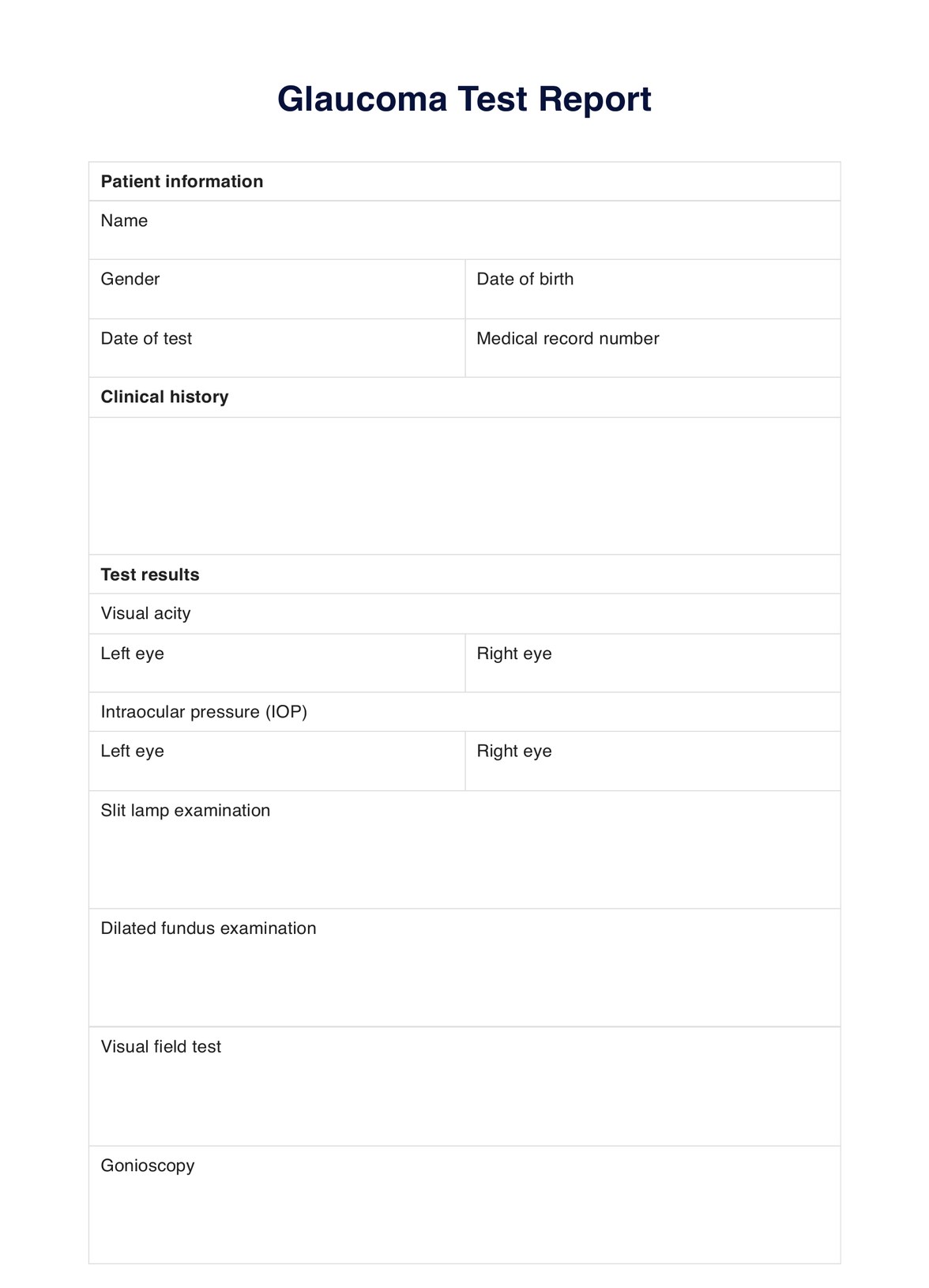
Visual acuity
Using an eye chart, the visual acuity test measures how well the patient can see at different distances. Results are typically recorded as a fraction, with 20/20 considered normal vision. A lower number indicates poorer vision, while a higher number indicates better vision.
Intraocular pressure (IOP)
The IOP test measures the pressure inside the eye, which can indicate glaucoma risk. Normal IOP ranges from 10 to 21 mmHg but can vary based on age and corneal thickness. Higher IOP may be an early sign of glaucoma but is not definitive and requires further testing.
Slit lamp examination
The slit lamp examination evaluates the anterior eye structures, including the cornea, iris, and lens. It helps detect abnormalities or signs of glaucoma. Results are recorded as normal or abnormal, with further testing if abnormalities are noted.
Dilated fundus examination
The dilated fundus examination allows viewing of the eye's interior, including the optic nerve and retina. It helps detect glaucoma-related damage, such as optic nerve thinning or changes in blood vessel appearance. Results are either normal or abnormal.
Visual field test
The visual field test assesses peripheral vision and detects abnormalities indicative of glaucoma. Results are typically plotted as a graph against normal values. Deviations from the norm may suggest potential vision loss due to glaucoma.
Gonioscopy
Gonioscopy evaluates the drainage angle of the eye where fluid flows out. It identifies blockages or abnormalities that may increase glaucoma risk. Results are recorded as open (normal), closed, or narrow-angle.
Optical coherence tomography (OCT)
OCT is a non-invasive imaging technique that produces high-resolution cross-sectional eye images using light waves. It detects changes in eye structures, such as optic nerve thinning or retinal layer changes indicative of glaucoma. Results are recorded as normal or abnormal.
Scanning laser polarimetry (SLP)
SLP measures the thickness of the retinal nerve fiber layer using light waves. It detects thinning or changes in this layer, indicating possible glaucoma. Results are recorded as within normal limits or outside normal limits.