
What is an Eye Physical Examination?
An eye exam, also known as an ophthalmic or optometric examination, is a comprehensive assessment of the visual health and function of the eyes. It involves a series of tests and evaluations performed by an eye care professional, such as an optometrist or ophthalmologist, to determine the overall condition of the eyes and identify any potential issues.
Various aspects of vision and eye health are scrutinized during an eye exam. The process typically includes measuring visual acuity, assessing refractive error (nearsightedness, farsightedness, or astigmatism), and examining the eye's internal and external structures, such as the cornea, lens, pupil, and iris eyelid of the left and right eyes.
An eye exam is not limited to individuals with vision problems. Those with normal vision should undergo periodic eye exams to maintain eye health. As many cases of vision loss and eye conditions (including certain glaucoma and macular degeneration types) are preventable, regular eye exams are key for early detection of potential problems.
A comprehensive physical examination of the eyes involves a series of assessments to evaluate various aspects of visual health and detect potential issues that may affect the eyes or vision. Here are some common components:
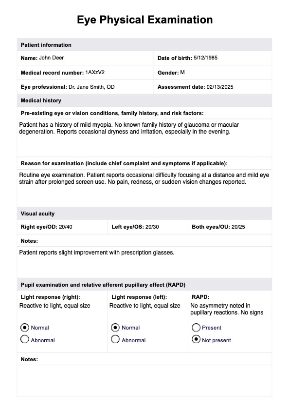
Visual acuity testing
Visual acuity testing measures the sharpness and clarity of vision, typically using a Snellen chart (where letters or symbols are read from a standardized distance). This assessment helps identify refractive errors such as myopia (nearsightedness), hyperopia (farsightedness), or astigmatism.
Pupils
This test involves assessing the size, equality, and response to light of the pupils. Relative afferent pupillary (RAPD) may be checked by shining a light into one pupil at a time and observing the constriction response in both pupils. This test provides valuable information about optic nerve function and potential neurological issues.
Extraocular motility and alignment
Evaluating extraocular motility and alignment assesses the coordination and movement of the eyes. It helps identify conditions such as strabismus (misalignment) and ensures proper eye teaming for binocular vision.
Intraocular pressure
This test is performed using a tonometer and is crucial for detecting glaucoma. Increased intraocular pressure can damage the optic nerve over time.
Confrontation visual fields
This test checks the patient's ability to see objects in their peripheral vision without directly looking at them. This test can reveal defects in the visual field, which may be indicative of various eye or neurological problems.
External examination
The external examination involves inspecting the eyelids, lashes, and surrounding tissues for abnormalities. Any signs of inflammation, infection, or other issues are carefully noted during this part of the exam.
Slit-lamp examination
The slit-lamp examination provides a detailed view of the anterior and posterior segments of the eye. It helps identify abnormalities in the cornea, lens, and other structures that may contribute to eye problems.
Fundoscopic examination
This technique typically involves stabilizing the patient's shoulder and using an ophthalmoscope to examine the interior of the eye, including the optic nerve, retina, and blood vessels. This is crucial for detecting conditions like diabetic retinopathy and macular degeneration.


