


What is posterolateral knee instability?
Posterolateral knee instability (PLKI) is a condition characterized by laxity or looseness in the posterolateral corner (PLC) of the knee, which can lead to significant stability issues and discomfort. This instability affects the outer aspect of the knee, where the tibia (shinbone) meets the femur (thighbone), compromising the knee's ability to withstand rotational forces.
Symptoms of posterolateral knee instability often include pain and swelling on the outside of the knee, a feeling of the knee giving way during activity, and difficulty with twisting or turning movements. Patients may also report a sensation of the knee locking or catching, particularly when attempting to change direction quickly.
Causes of posterolateral knee instability
Posterolateral knee instability often results from traumatic injuries, typically seen in sports or severe accidents, where the knee experiences direct blows or undergoes sudden, forceful twists, potentially damaging the anterior cruciate ligament and other critical structures for lateral knee stability.
Injuries to the lateral ligament compartment, including the lateral collateral ligament, the popliteofibular ligament, and the posterolateral corner injury, are common sources of instability. These structures are vital for preventing excessive lateral opening and rotational instability of the knee. Isolated fibular collateral ligament injuries, although less common, also significantly contribute to overall instability.
Combined posterolateral knee injuries pose a particular challenge as they affect multiple stabilizing components simultaneously, often requiring comprehensive approaches including posterior cruciate ligament reconstruction to restore stability and function adequately. These complex injuries require a thorough understanding of knee mechanics and often involve multiple surgical interventions to adequately restore stability and function.
How to diagnose a posterolateral knee instability or injury?
Diagnosing PLKI often starts with a detailed medical history and physical examination, focusing on any incidents that could have led to knee injuries. Key diagnostic tools include:
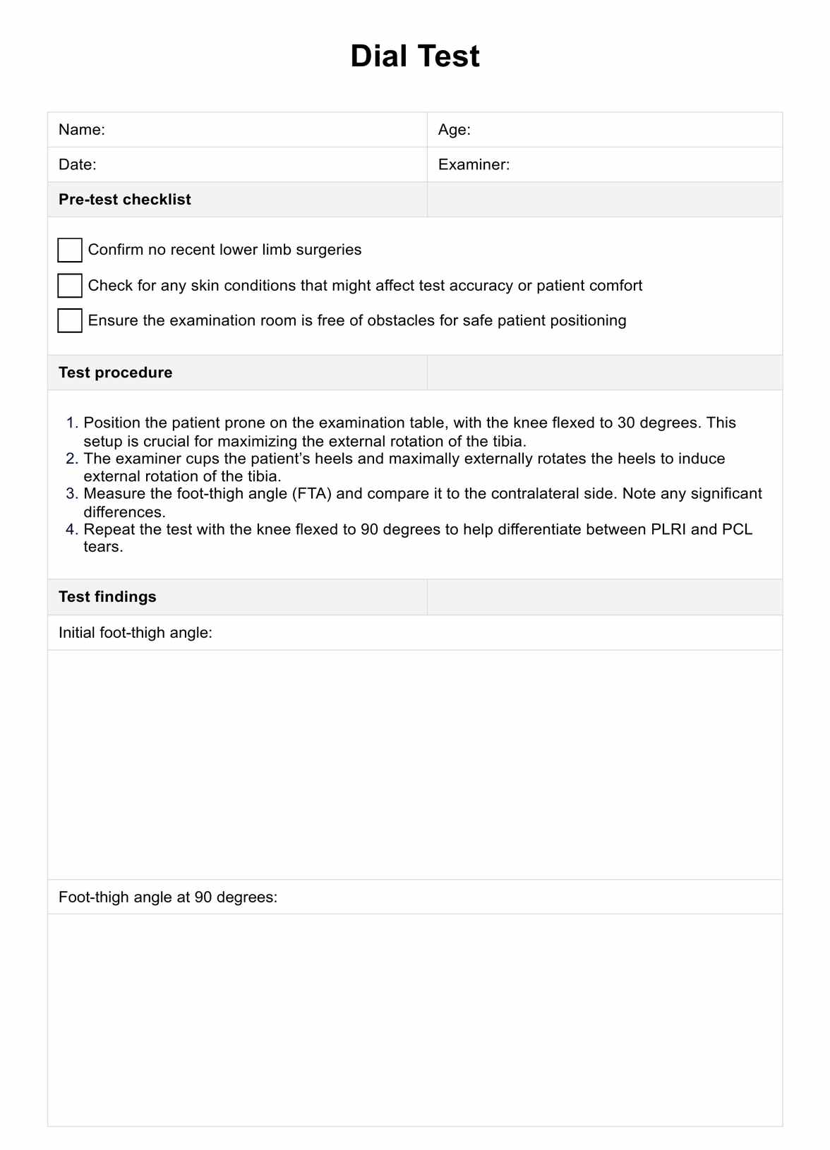
- Physical tests: Specific maneuvers, like the Dial Test, assess the integrity of the PLC.
- Imaging: MRI scans are particularly useful for visualizing the extent of soft tissue damage, while X-rays help to rule out bone fractures.
- Stress radiography: This can be used to assess the degree of knee laxity more precisely.
Proper diagnosis is crucial as it guides the subsequent treatment and rehabilitation processes, ensuring the best possible recovery and return to function for the patient.


