Circle of Willis Diagram

Download a free Circle of Willis diagram and better understand its function, structure, and significance in the brain's circulatory system.

By Matt Olivares on Feb 02, 2025.

tick

Fact Checked by Ericka Pingol.

Use Template
AI IconToolbarShare ui

What is the circle of Willis?

The circle of Willis (Circulus arteriosus cerebri) is a vascular structure located at the base of the brain that links the internal carotid and vertebrobasilar arterial systems, supplying consistent blood flow between the brain's anterior and posterior arteries. By forming a collateral pathway, it minimizes the risk of ischemia by redirecting blood flow in the event of arterial blockages. Its typical structure includes the anterior, middle, and posterior cerebral arteries (PCAs) connected by the anterior and posterior communicating (PCOM) arteries. Intracranial aneurysms occur when there is a weakening in one of the blood vessel walls, often in the Circle of Willis, making it susceptible to rupture.

Surrounding the stalk of the pituitary gland, the circle of Willis establishes critical connections between the blood supplies of the forebrain and the hindbrain. This connection bridges the internal carotid and vertebrobasilar systems, compensating for the loss of primitive embryonic vascular links (Krabbe-Hartkamp, 1998; Standring, 2021).

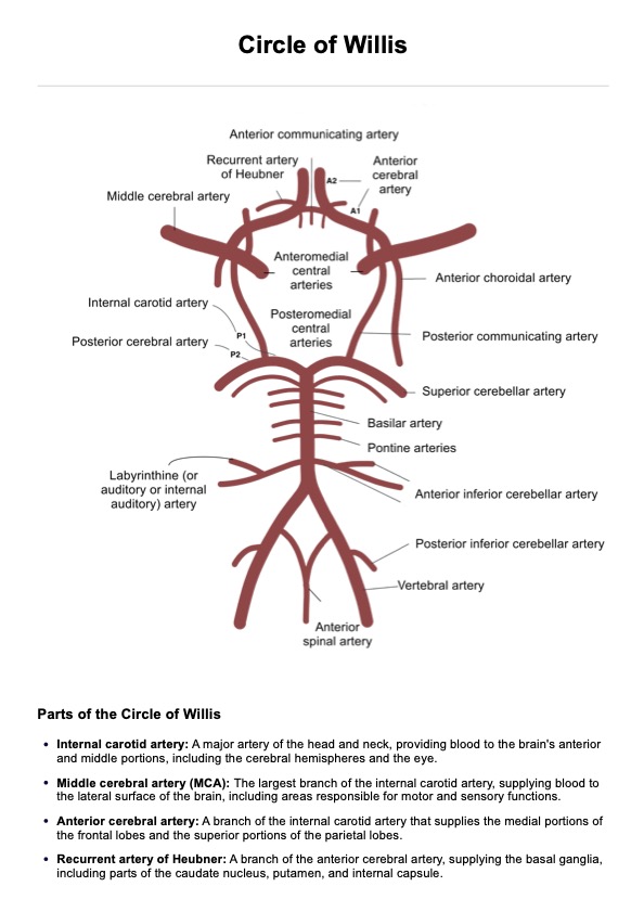
The primary components of the circle of Willis are:

  • Anterior cerebral arteries (ACAs): Paired arteries arising from the internal carotid arteries (ICAs).
  • Anterior communicating artery (ACOM): A single midline artery connecting the ACAs.
  • Internal carotid arteries (ICAs): Branches of the common carotid arteries.
  • Posterior cerebral arteries (PCAs): Paired arteries originating from the basilar artery (BA).
  • Posterior communicating (PCOM) arteries: Paired arteries linking the ICAs and PCAs.

The anterior circulation comprises the ACAs, ACOM, and middle cerebral arteries (MCAs), which branch from the ICAs. The posterior circulation includes the PCAs, originating from the basilar artery formed by the vertebral arteries. If one of our brain's significant arteries becomes narrow (which slows down blood flow) or there is a blockage (blood flow stops), the Circle of Willis will work to maintain blood circulation by redirecting the blood flow to interconnected branches. This redirection is called collateral blood flow, which means alternative routes will be used if blood flow is disrupted due to an artery being damaged, narrow, or blocked.

What will you see in a Circle of Willis Diagram?

The Circle of Willis Diagram allows you to see the different components compromising the circle of Willis visually. The key features are:

  • Internal carotid artery: Supplies blood to the anterior and middle portions of the brain, including the cerebral hemispheres and eye.
  • Middle cerebral artery (MCA): The largest branch of the internal carotid artery, delivering blood to the brain's lateral surface, including motor and sensory areas.
  • Anterior cerebral artery: Branch of the internal carotid artery, providing blood to the frontal and superior parietal lobes.
  • Recurrent artery of Heubner: Branch of the anterior cerebral artery, supplying the basal ganglia and parts of the caudate nucleus and putamen.
  • Anterior communicating artery: Connects the left and right anterior cerebral arteries, contributing to the Circle of Willis.
  • Posteromedial central arteries: Small branches from the posterior cerebral artery, supplying the occipital lobe, thalamus, and brainstem.
  • Posterior cerebral artery: Arises from the basilar artery, supplying the occipital lobe, inferior temporal lobe, and thalamus.
  • Posterior communicating artery: Connects the posterior cerebral artery with the internal carotid artery, completing the Circle of Willis.
  • Superior cerebellar artery: Branch of the basilar artery, supplying the superior cerebellum and midbrain.
  • Pontine arteries: Branches of the basilar artery, supplying the pons.
  • Basilar artery: Formed by the union of the vertebral arteries, supplying the brainstem, cerebellum, and occipital lobe.
  • Anterior inferior cerebellar artery: Branch of the basilar artery, supplying the anterior and inferior cerebellum and pons.
  • Labyrinthine artery: Supplies the inner ear, including the cochlea and vestibular system.
  • Posterior inferior cerebellar artery: Branch of the vertebral artery, supplying the posterior cerebellum and medulla.
  • Vertebral artery: Arises from the subclavian artery, supplying the posterior brain, including the brainstem and cerebellum.
  • Anterior spinal artery: Formed by branches from the vertebral arteries, supplying the anterior spinal cord.

To access our free Circle of Willis Diagram, click the "Use template" button, which lets you open the handout via the Carepatron app. For a PDF copy, click "Download."

When is it best to use a Circle of Willis Diagram?

To help people visualize the arteries' arrangement, we created a diagram of the Circle of Willis. This diagram shows how the arteries are distributed and which brain regions they connect to. This diagram is useful for neurologists and doctors as both a reference and an educational tool. For those who need a visual aid, the diagram can be printed and displayed in offices or study areas as a reminder of the brain's arterial structure.

The diagram is also great for educational purposes. For instance, if you're creating a neurology textbook or teaching, you can include the diagram alongside explanations of each part. Additionally, it can be printed and handed out to students or used in PowerPoint presentations during lectures, offering a helpful tool for note-taking and review during exam preparation.

What are the benefits of using a Circle of Willis Diagram?

Here are the benefits of using our free Circle of Willis template:

Visual understanding

The diagram provides a clear representation of the Circle of Willis, helping to understand the arrangement and connections of the arteries in the brain. It simplifies complex anatomical details and allows for easier comprehension of how blood flows to different regions of the brain.

Educational and study aid

The diagram serves as both an educational tool for teaching and a study aid for students. It helps students and professionals alike grasp the key concepts of brain vascular anatomy. In a classroom setting, it can be used to explain the structure, while students can use it to take notes, review, and prepare for exams.

Quick reference

For medical professionals, the diagram is a quick reference to refresh knowledge about the brain’s arterial system. It’s a valuable resource in clinical settings, where understanding the Circle of Willis can be crucial for diagnosis and treatment planning.

Patient and family education

The diagram can also be a helpful tool for explaining medical conditions related to the Circle of Willis to patients and their families. By visually showing how blood flows through the brain, it can aid in discussions about stroke risk, vascular health, and treatment options, making complex information easier to understand.

References

Krabbe-Hartkamp, M. J., Van der Grond, J., De Leeuw, F. E., De Groot, J. C., Algra, A., Hillen, B., ... & Mali, W. P. (1998). Circle of Willis: morphologic variation on three-dimensional time-of-flight MR angiograms. Radiology, 207(1), 103-111.

Standring, S. (Ed.). (2021). Gray’s anatomy: The anatomical basis of clinical practice (42nd ed.). Elsevier.

What happens if the circle of Willis is blocked?

Commonly asked questions

What happens if the circle of Willis is blocked?

If the circle of Willis is blocked, it can often compensate by rerouting blood flow through alternate pathways. However, if this collateral circulation is insufficient, it may lead to reduced blood supply to parts of the brain, causing ischemia or a stroke.

What is the circle of Willis and what does it do?

The circle of Willis is a ring of interconnected arteries at the base of the brain. It ensures consistent blood flow by rerouting blood if a blockage or narrowing occurs in one of the main arteries supplying the brain.

What is the most common anomaly of the circle of Willis?

The most common anomaly is hypoplasia (underdevelopment) of one or both posterior communicating arteries (PCOMs). This can affect the circle’s ability to provide collateral blood flow.

Join 10,000+ teams using Carepatron to be more productive

One app for all your healthcare work