How to use the Peripheral Vascular Examination template
This template is a valuable guide to conducting a clinical evaluation of blood flow and blood pressure abnormalities. Follow these steps to properly assess signs of peripheral vascular disease or peripheral artery disease pathology:
Step 1: Access the template
Open the template by clicking the "Use template" button on this page, which opens the template in the Carepatron app. From there, you can customize the template before filling or printing it out.
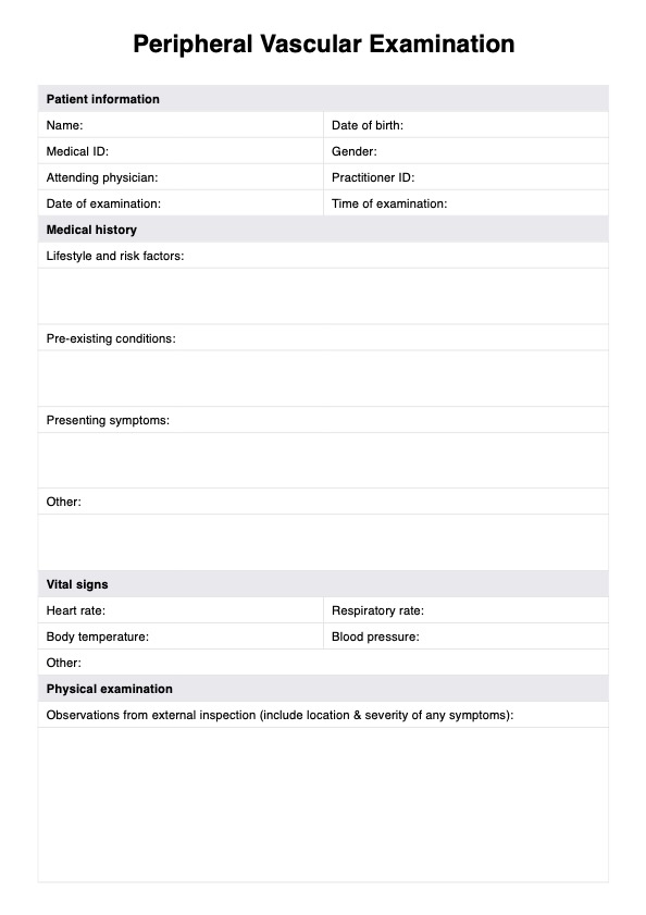
Step 2: Collect patient history
Input the patient's key demographic information at the top of the form. Collect relevant details around the patient's lifestyle, medical history, leg pain (which can indicate ischemia due to reduced blood flow), and any other symptoms. If the patient is diabetic, ask about their foot care and any history of foot ulcers.
Step 3: Vital signs and external examination
The initial stage of the physical assessment involves checking and recording the patient's vital signs. Examine the legs for signs of pitting edema, varicose veins, and venous ulcers that tend to appear on the medial aspect of the leg. If arterial ulcers are present (typically found on the foot or near the medial malleolus), record these also.
Step 4: Pulse examination
Next, palpate pulses in the legs (such as the radial pulse) and upper extremities to assess arterial flow. Absent pulses or diminished pulses may suggest artery disease or a blood clot. Also check and record capillary refill time.
Step 5: Blood pressure measurement
Perform the ankle brachial index (ABI) and record blood pressure readings of the arms and legs. A significant difference may indicate peripheral arterial pathology. In some cases it may also be appropriate to perform further tests such as the Doppler ultrasound to assess blood flow and blood pressure in the peripheral arteries. This identifies areas of reduced flow that might suggest the need for vascular surgery or stent placement. Record the results.
Step 6: Imaging tests
Detailed imaging of the arteries, including the renal arteries and the aorta, can identify specific areas of stenosis or blockages. If further imaging is performed, record the tests and their findings in the template.
Step 7: Diagnosis and treatment
Disclose the findings of the assessment (including any diagnoses, signs of disease progression, or identified risk factors) to the patient. If necessary, work with the patient to devise a treatment plan. This may include supervised exercise therapy, prescriptions, referrals, or advice on lifestyle changes to manage risk factors (such as smoking cessation, managing diabetes, and supervised exercise therapy).
Step 8: Monitoring and follow-up
Regular follow-up appointments are crucial for monitoring the disease's progression, evaluating the treatment plan's effectiveness, and making adjustments as needed. This may include repeat vascular testing and reassessment of cardiovascular risk factors.