What is a PVD Test?

A PVD test, or peripheral vascular disease test, is a diagnostic procedure used to evaluate blood flow and blood pressure in the blood vessels, particularly in the limbs, to diagnose conditions such as peripheral artery disease (PAD). 

This test helps identify risk factors and symptoms of poor blood flow, such as leg pain or intermittent claudication. It involves various noninvasive methods like ankle-brachial index (ABI) measurement, Doppler ultrasound flow studies, pulse volume recording (PVR), magnetic resonance angiography (MRA), and physical examination.

During the PVD test, blood pressure measurements are taken at different points in the limbs using a blood pressure cuff and Doppler ultrasound to assess blood flow. An ABI test compares blood pressure readings in the ankle and arm to detect reduced blood flow. Additionally, sound waves are used to evaluate blood vessel abnormalities and the presence of narrowed arteries or blood clots.

Medical and family history, as well as other risk factors such as high blood pressure, high cholesterol, diabetes, and smoking, are considered during the evaluation. Lifestyle changes like regular exercise, quitting smoking, and managing cholesterol levels are often recommended to prevent peripheral vascular disease and improve blood circulation. 

If severe PVD is detected, further treatment options such as balloon angioplasty, bypass surgery, or vascular surgery may be necessary to restore blood flow and prevent complications like heart attacks or limb ischemia.

Printable PVD Test

Download this PVD Test for accurate assessment and diagnosis of peripheral vascular disease, enabling prompt intervention and personalized treatment plans for optimal patient care.

Why would a doctor recommend a PVD Test?

A doctor may recommend a PVD test, or peripheral vascular disease test, for several reasons:

  • Presence of symptoms: If a patient experiences symptoms suggestive of peripheral artery disease (PAD) or peripheral vascular disease (PVD), such as leg pain, cramping, numbness, or weakness, particularly during physical activity (intermittent claudication), a doctor may order a PVD test to confirm the diagnosis and assess disease severity.
  • Risk factors: Individuals with risk factors for PVD, such as advanced age, smoking, diabetes, high blood pressure, high cholesterol levels, obesity, or a family history of vascular disease, may undergo screening tests to detect early signs of vascular abnormalities or reduced blood flow.
  • Evaluation of known disease: For patients already diagnosed with PAD or PVD, regular monitoring with PVD tests helps track disease progression, assess treatment effectiveness, and detect complications like blood clots or narrowed arteries.
  • Preventive screening: In some cases, especially for individuals with multiple risk factors or a family history of vascular disease, doctors may recommend PVD testing as part of preventive healthcare to identify early signs of reduced blood flow and initiate interventions to prevent disease progression or complications.
  • Preoperative assessment: Before specific surgical procedures, such as vascular surgeries or limb amputations, doctors may order PVD tests to evaluate blood flow and ensure optimal surgical planning and outcomes.

How is a PVD Test performed?

A PVD test, or peripheral vascular disease test, involves several noninvasive procedures to evaluate blood flow and blood pressure in the limbs. The specific tests may vary depending on the patient's symptoms, medical history, and the doctor's recommendations. Here's an overview of commonly used methods in a PVD test:

  • Physical examination: The doctor will start by conducting a physical exam to assess symptoms, check pulses in various locations of the body, and examine the skin for signs of poor circulation, such as ulcers or discoloration.
  • Medical and family history: The doctor will inquire about the patient's medical history, including any existing conditions such as diabetes, high blood pressure, high cholesterol, or previous vascular problems. A family history of vascular diseases is also crucial in assessing genetic predispositions.
  • Ankle-brachial index (ABI) test: This test compares blood pressure measurements taken at the ankle and arm using a blood pressure cuff and Doppler ultrasound. A lower ankle pressure than the arm indicates reduced blood flow to the legs, suggesting peripheral artery disease (PAD).
  • Pulse volume recording (PVR): PVR measures changes in blood volume in the limbs using pressure cuffs and sensors. It provides information about blood flow patterns and detects abnormalities such as blockages or narrowed arteries.
  • Doppler ultrasound: Doppler ultrasound uses sound waves to create images of blood flow in the arteries and veins. It helps identify the location and severity of blockages, narrowing, or blood clots in the blood vessels.
  • Magnetic resonance angiography (MRA): MRA is a noninvasive imaging technique that uses magnetic fields and radio waves to visualize blood vessels in detail. It provides high-resolution images of the arteries and can detect abnormalities such as narrowed or aneurysms.
  • Treadmill exercise test: In some cases, a treadmill exercise test may be performed to assess symptoms of intermittent claudication (leg pain during walking). The test measures how far the patient can walk on a treadmill before experiencing symptoms, helping to gauge the severity of peripheral artery disease.
  • Additional tests: Depending on the findings and clinical suspicion, tests such as blood tests (to check cholesterol levels, blood sugar, etc.), CT angiography, or angiography may be recommended for further evaluation.

Throughout the PVD test, the patient remains awake without anesthesia. These noninvasive tests are generally safe and well-tolerated, providing valuable information to diagnose peripheral vascular disease, assess blood flow, and guide treatment decisions.

Preparations needed before undergoing a PVD Test

Before undergoing a PVD test (Peripheral Vascular Disease test), specific preparations may be necessary to ensure accurate results and a smooth testing process. Here's a general guide on what to expect and how to prepare:

  • Follow instructions from healthcare provider: Your healthcare provider will provide specific instructions tailored to your needs and the type of PVD test being performed. Follow these instructions carefully to ensure accurate results.
  • Medication review: Inform your healthcare provider about all medications you currently take, including prescription medications, over-the-counter drugs, vitamins, and supplements. Some medicines may need to be adjusted or temporarily stopped before the test.
  • Fasting: Depending on the type of PVD test being performed, you may be instructed to fast for a certain period before the test. This is particularly important for tests that involve blood work or imaging techniques that require clear images without interference from food or drink.
  • Comfortable clothing: Wear loose, comfortable clothing that allows easy access to the limbs being tested. Avoid wearing tight clothing, as it may interfere with blood pressure measurements or other aspects of the test.
  • Stay hydrated: Drink plenty of water before the test unless instructed otherwise by your healthcare provider. Staying hydrated helps ensure accurate blood pressure measurements and facilitates the testing process.
  • Avoid smoking and caffeine: If you are a smoker, try to avoid smoking for several hours before the test, as smoking can temporarily affect blood flow and blood pressure readings. Similarly, avoid consuming caffeinated beverages like coffee or tea, as caffeine can also influence blood pressure.
  • Bring necessary documents: Bring any relevant medical records, such as your medical history, list of medications, and insurance information, to the appointment. This information will help your healthcare provider assess your overall health and plan the appropriate PVD test.
  • Arrive on time: Arrive at the testing facility on time for your scheduled appointment. This allows ample time for registration, paperwork, and any necessary preparations before the test.
  • Inform about allergies or medical conditions: Inform your healthcare provider about any allergies or medical conditions you are currently experiencing. This information helps ensure your safety during the test and allows for appropriate precautions to be taken if needed.

How does our PVD Test template work?

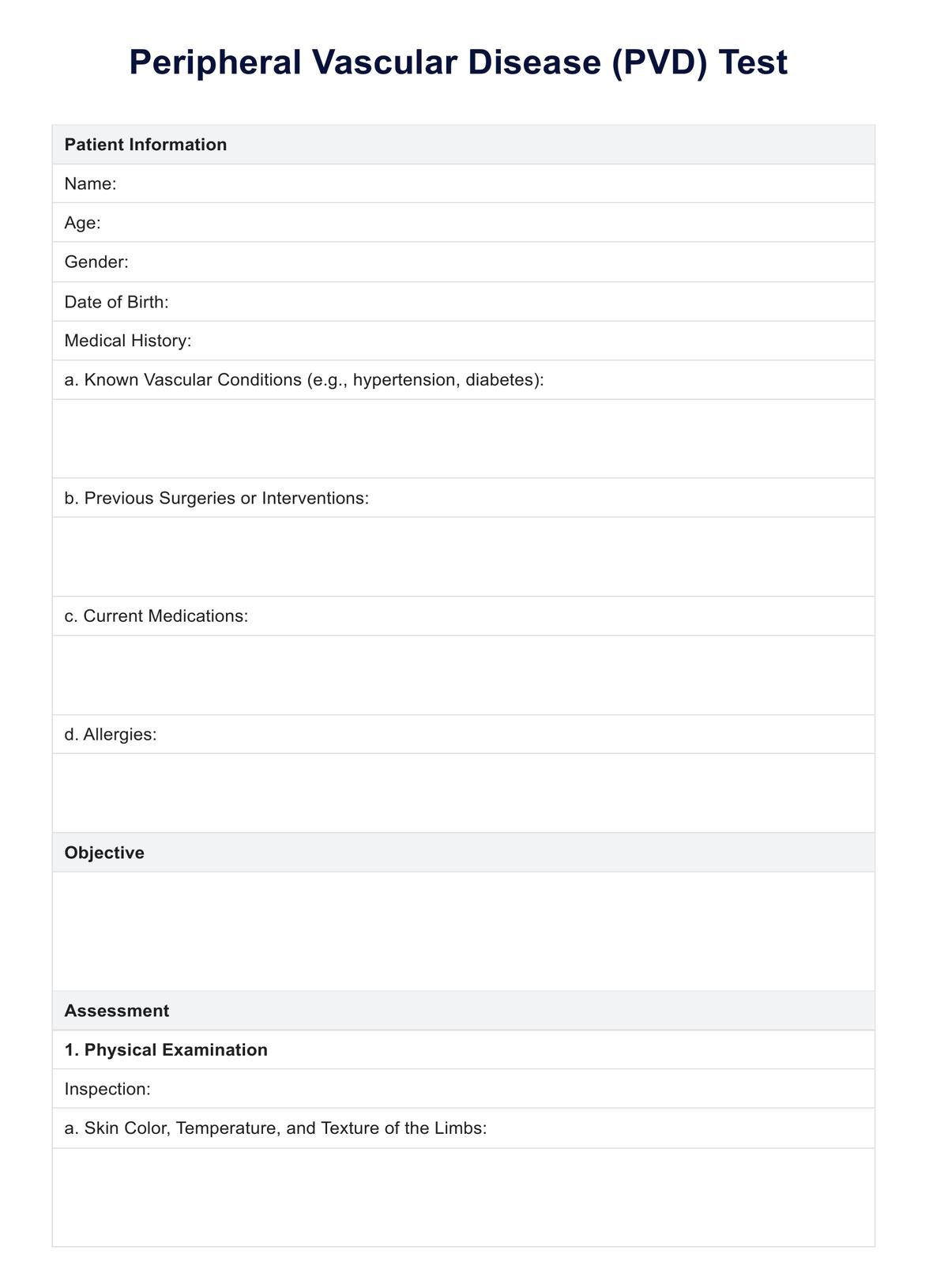
The PVD Test template is designed to guide medical professionals through assessing, diagnosing, and managing patients suspected of having Peripheral Vascular Disease (PVD). Here's how it works:

  1. Patient information: This section captures essential demographic details and medical history relevant to the assessment of PVD. It includes the patient's name, age, gender, date of birth, known vascular conditions, previous surgeries or interventions, current medications, and allergies.
  2. Objective: The objective section outlines the purpose of the evaluation: to assess the vascular status and perfusion of the patient's limbs using various diagnostic tests for PVD.
  3. Assessment: This section breaks down the assessment process into three main categories: physical examination, laboratory tests, and diagnostic imaging.some text
    • Physical examination: This involves inspecting the patient's limbs for visual signs of PVD, palpating pulses, assessing capillary refill time, auscultating for bruits, and performing additional tests such as Ankle-Brachial Index (ABI) measurement and Doppler ultrasound examination.
    • Laboratory tests: This includes ordering relevant blood tests such as complete blood count (CBC), lipid profile, and blood glucose levels to assess cardiovascular risk factors and rule out other conditions.
    • Diagnostic imaging: This section outlines the use of imaging modalities like duplex ultrasound, magnetic resonance angiography (MRA), computed tomography angiography (CTA), and digital subtraction angiography (DSA) to visualize arterial structures and assess blood flow.
  4. Interpretation: Based on the findings from the assessment, the medical professional interprets the results to determine the presence, severity, and location of PVD.
  5. Plan: This section outlines the management plan tailored to the patient's condition, which may include lifestyle modifications, pharmacological therapy, interventional procedures, referrals, and follow-up appointments.
  6. Patient education: The template emphasizes the importance of patient education regarding PVD, its risk factors, treatment options, and signs/symptoms requiring immediate medical attention.
  7. Documentation: It emphasizes accurate documentation of assessment findings, diagnostic results, management plans, patient education, and signatures for medical records.

Why use Carepatron as your cardiology software?

Carepatron is an ideal choice for cardiology software due to its comprehensive features tailored to the specific needs of healthcare professionals, including cardiologists. The platform's HIPAA compliance ensures the secure handling of sensitive patient information, which is crucial in cardiology, where patient data privacy is paramount. 

By offering appointment reminders, we effectively reduce the number of no-shows and last-minute cancellations, optimizing the scheduling process for cardiology practices. Moreover, the online scheduling feature allows patients to book appointments conveniently, enhancing accessibility and patient satisfaction.

Our integration capabilities with clinical documentation, telehealth video conferencing, and medical billing streamline cardiology practices, maximizing efficiency and quality of care. The software's medical dictation solution, with advanced security measures like high-level encryption, ensures the safe storage and transmission of patient data, a critical aspect in cardiology, where data security is essential.

medical dictation solution
What is the PVD Test?

Commonly asked questions

What is the PVD Test?

PVD testing involves assessing the vascular status and perfusion of the limbs to diagnose Peripheral Vascular Disease, typically through physical examination, vascular imaging, and noninvasive tests like the Ankle-Brachial Index (ABI).
What is the screening for PVD?

Screening for PVD often includes measuring the Ankle-Brachial Index (ABI), which compares blood pressure in the ankle to that in the arm. Physical examination for pulse abnormalities and risk factor assessment also play vital roles.

How do you examine PVD?

Examination for PVD involves inspecting skin color, temperature, and texture, palpating peripheral pulses, assessing capillary refill time, auscultating for bruits, and performing noninvasive tests like ABI measurement and Doppler ultrasound.

Join 10,000+ teams using Carepatron to be more productive

One app for all your healthcare work