Gout Diagnosis Criteria

Uncover the essential Gout Diagnosis Criteria here, including symptoms, diagnostic tests, and the American College of Rheumatology guidelines.

By Nate Lacson on Apr 10, 2024.

Fact Checked by Ericka Pingol.

Use Template

What is gout?

Gout, also known as gouty arthritis, is a common form of inflammatory arthritis characterized by sudden, severe attacks of pain, swelling, redness, and tenderness in the joints. Gout typically affects the joint at the base of the big toe, but it can also occur in other joints such as the ankles, knees, wrists, fingers, and elbows.

The condition is caused by the accumulation of urate crystals in the joints, resulting from elevated levels of uric acid in the blood. Uric acid is a waste product formed from the breakdown of purines, which are substances found in various foods and in the body's cells. When the body produces too much uric acid or the kidneys excrete too little, uric acid can build up and form needle-like crystals in the joints, triggering the inflammatory response seen in gout.

Acute gout attacks often occur suddenly, often at night, and can be triggered by factors such as alcohol consumption, high-purine foods, stress, and certain medications. Chronic gout (also known as chronic gouty arthritis), on the other hand, involves repeated episodes of pain and inflammation, which can eventually lead to joint damage and tophi, which are lumps of urate crystals that can form under the skin.

The American College of Rheumatology, publisher of the journal Arthritis & Rheumatology (Arthritis Rheum), provides guidelines for diagnosing and managing gout. According to their criteria, a gout diagnosis can be confirmed by identifying urate crystals in joint fluid or the presence of a tophus.

The prevalence of gout varies by region and demographic factors, with the condition being more common in men than in women and increasing in frequency with age. Risk factors for developing gout include genetics, obesity, hypertension, kidney disease, and certain dietary habits.

Symptoms of gout

Gout symptoms can vary depending on the stage of the condition. In the early stages, known as acute gout or acute gouty arthritis, symptoms typically come on suddenly and can include:

  • Intense joint pain: Gout usually affects the large joint of the big toe but can occur in any joint. The pain is most severe within the first four to twelve hours after it begins.
  • Lingering discomfort: After the severe pain subsides, some joint discomfort may last from a few days to a few weeks. Later attacks are likely to last longer and affect more joints.
  • Inflammation and redness: The affected joint or joints become swollen, tender, warm, and red.
  • Limited range of motion: As gout progresses, you may not be able to move your joints normally.

In the later stages, known as chronic gout, the symptoms can become more persistent and can include:

  • Chronic joint pain: The pain and inflammation can become chronic, affecting more than one joint.
  • Tophi: The formation of tophi, which are hard, uric acid crystals deposits under the skin, can occur. Tophi can develop in various areas such as the fingers, toes, elbows, or ears.
  • Joint damage: Untreated gout can lead to joint damage and deformity.

How is this different from arthritis?

Gout is often classified as a type of arthritis due to the inflammation it causes in the joints. However, it is distinct from other forms of arthritis in several ways:

  • Gout (acute gouty arthritis): Caused by the accumulation of uric acid crystals in the joints, leading to intense pain and swelling, typically in one joint at a time. Attacks are sudden and severe, often occurring at night.
  • Rheumatoid arthritis: An autoimmune disorder causing chronic inflammation of multiple joints, with persistent symptoms that develop gradually.
  • Osteoarthritis: Characterized by the gradual wear and tear of joint cartilage, without the formation of uric acid crystals.
  • Septic arthritis (acute arthritis): Caused by an infection in the joint, leading to pain, swelling, and fever. This condition requires immediate medical attention.

Understanding the differences between these types of arthritis is crucial for accurate diagnosis and effective treatment.

Primary gout vs. secondary gout

Primary gout, also known as idiopathic gout, is the most common form and occurs when the body produces too much uric acid or fails to excrete it properly. This imbalance can be due to genetic factors, lifestyle choices such as diet and alcohol consumption, or the natural aging process.

In primary gout, the kidneys may be healthy but are unable to keep up with the excess uric acid, leading to its accumulation and crystal formation in the joints. The management of primary gout focuses on long-term lifestyle changes and medication to control uric acid levels and prevent future attacks.

Secondary gout, on the other hand, results from other medical conditions or treatments that increase uric acid levels. For example, certain cancers, blood disorders, and kidney diseases can lead to secondary gout. Medications such as diuretics and low-dose aspirin can also contribute to its development.

In these cases, treating the underlying condition or adjusting medication can often alleviate the gout symptoms. Understanding the distinction between primary and secondary gout is essential for healthcare professionals to provide targeted and effective treatment, addressing both the symptoms and the root cause of the elevated uric acid levels.

Printable Gout Diagnosis Criteria

Download this Gout Diagnosis Criteria for precise identification and swift treatment, enhancing patient care.

How do healthcare professionals diagnose gout?

Diagnosing gout involves a comprehensive evaluation by healthcare professionals to distinguish it from other conditions that may present with similar symptoms, such as rheumatoid arthritis or pseudogout. The diagnostic process can be dividend into three parts, each with its own smaller parts:

1. Clinical evaluation

A thorough clinical evaluation is the first step in diagnosing gout. This includes assessing the patient's medical history and conducting a physical examination. Here's what this evaluation entails:

  • Medical history: The healthcare professional will inquire about the patient's symptoms, duration of attacks, frequency of episodes, and any family history of gout.
  • Physical examination: The physician will examine the affected joints for signs of inflammation, swelling, redness, and tenderness.

2. Laboratory tests

Laboratory tests are crucial for confirming the presence of high uric acid levels and identifying urate crystals in the joint fluid. Here are the relevant lab tests:

  • Blood test: A blood sample may be taken to measure the levels of uric acid in the blood. High uric acid levels are associated with gout, but they can also be elevated in people without gout or normal during a gout attack.
  • Synovial fluid analysis (joint fluid analysis): If the diagnosis is uncertain, a healthcare professional may use a needle to draw fluid from the affected joint (joint aspiration) and examine it for urate crystals under a microscope. The presence of urate crystals confirms a diagnosis of gout.

3. Imaging tests

Imaging tests play a supportive role in diagnosing gout by visualizing joint changes and detecting urate crystals. These tests may be done:

  • X-rays: While X-rays cannot detect gout in the early stages, they can help rule out other causes of joint inflammation and assess any joint damage in chronic gout.
  • Ultrasound: This imaging technique can detect urate crystals in joints, even when there are no visible signs of a gout attack.
  • Dual-energy CT scan: This specialized CT scan can detect the presence of urate crystals in joints, even when not flaring, but it is not commonly used for diagnosing gout due to its high cost.

What are the diagnostic criteria for gout?

To accurately diagnose gout, healthcare professionals rely on specific clinical diagnostic criteria. The American College of Rheumatology (ACR) and the European League Against Rheumatism (EULAR) have jointly developed the ACR/EULAR Gout Classification Criteria which serve as both diagnostic and classification criteria for gout.

These criteria are the new gout classification criteria for clinical studies and research rather than for diagnosing individual patients, but they can still provide valuable guidance for healthcare professionals.

ACR/EULAR gout classification criteria

The ACR/EULAR criteria for gout classification are based on a scoring system that includes clinical, laboratory, and imaging findings, similar to the approach used in systemic sclerosis classification criteria. The criteria below are used when a patient meets the entry criterion of having at least one episode of swelling, pain, or tenderness in a peripheral joint or bursa. A score of 8 or more is classified as gout.

  • Entry criterion: This is the preliminary criteria; the other criteria do not apply if this is not fulfilled. There should be at least one episode of swelling, pain, or tenderness in a peripheral joint or bursa.
  • Sufficient criterion: Check if monosodium urate (MSU) crystals exist in a symptomatic joint or bursa (i.e., in synovial fluid) or tophus. If one of these is present, it can be classified as gout without applying further criteria.

Additional Criteria

These are used if the Sufficient Criterion is not met. Each has an associated score. A total score greater or equal to 8 is required for classification as gout.

Clinical

  1. Pattern of joint/bursa involvement during symptomatic episode(s):
  • Joint(s) or bursa(e) other than ankle, midfoot, or 1st MTP: 0 points
  • Ankle OR midfoot (as part of monoarticular or oligoarticular episode without MTP1 involvement): 1 point
  • MTP1 (as part of monoarticular or oligoarticular episode): 2 points
  1. Characteristics of symptomatic episode(s):
  • Erythema overlying affected joint: 1 point
  • Can't bear touch or pressure to affected joint: 1 point
  • Great difficulty with walking or inability to use affected joint: 1 point
  1. Time-course of episode(s):
  • Presence of ≥2 typical episodes (time to maximal pain <24 hours, resolution of symptoms in ≤14 days, complete resolution between episodes): 2 points
  1. Clinical evidence of tophus: 4 points

Laboratory

  1. Serum urate concentration:
  • <4 mg/dL: -4 points
  • 4-<6 mg/dL: 0 points
  • 6-<8 mg/dL: 2 points
  • 8-<10 mg/dL: 3 points
  • ≥10 mg/dL: 4 points
  1. Synovial fluid analysis:
  • MSU negative: -2 points

Imaging

  1. Imaging evidence of urate deposition in symptomatic joint or bursa (ultrasound evidence of double-contour sign or DECT demonstrating urate deposition): 4 points
  2. Imaging evidence of gout-related joint damage (conventional radiography demonstrating at least one erosion): 4 points

How to use our Gout Diagnosis Criteria template

The Gout Diagnosis Criteria template is based on the ACR/EULAR Gout Classification Criteria and is designed to assist healthcare professionals in diagnosing gout effectively. Here are the steps to follow:

Step 1: Access and explain the template

Download the Gout Diagnosis Criteria template from our platform and explain its purpose and structure to the patient. This helps in setting the right expectations and understanding the importance of the information being collected.

Step 2: Fill in patient information and evaluate clinical features

Enter the patient's basic information such as name, date of evaluation, gender, and age. Then, review and check the boxes corresponding to the clinical features the patient exhibits, including the pattern of joint involvement and characteristics of symptomatic episodes.

Step 3: Record laboratory and imaging test results

Input the results of laboratory tests, particularly the serum urate concentration, and assess any imaging findings for evidence of urate deposition and gout-related joint damage.

Step 4: Confirm diagnosis

Based on the ACR/EULAR criteria and the total score, determine whether the patient can be classified as having gout. Record any additional notes that may assist in the patient's treatment plan.

Gout Diagnosis Criteria example (sample)

To assist healthcare professionals in the diagnosis and management of gout, Carepatron has developed a sample Gout Diagnosis Criteria template. The template is based on the ACR/EULAR Gout Classification Criteria and includes fields for clinical features, laboratory tests, imaging findings, and diagnostic confirmation.

Download our free Gout Diagnosis Criteria template example here.

Gout Diagnosis Criteria example (sample)

Why use Carepatron as your general practice software?

epatron is an all-in-one practice management software designed to streamline the operations of general practices. Here's why healthcare professionals should consider using Carepatron for their practice:

  • Integrated patient management: Carepatron offers comprehensive patient management features, including secure storage of patient records, easy appointment scheduling, and automated reminders. This ensures that patient information is organized and accessible, enhancing the efficiency of patient care.
  • Telehealth capabilities: With the increasing demand for remote consultations, Carepatron's telehealth feature allows healthcare professionals to conduct virtual appointments seamlessly. This ensures continuity of care while maintaining the safety and convenience of both patients and practitioners.
  • Streamlined billing and payments: Carepatron simplifies the billing process with automated invoicing and integrated payment processing. This reduces administrative burden and ensures timely payment for services rendered.
  • Collaborative care: The platform facilitates collaboration among healthcare professionals within a practice, enabling seamless communication and coordination of patient care. This fosters a team-based approach to healthcare, improving patient outcomes.

By incorporating Carepatron into their practice, healthcare professionals can enhance their workflow, improve patient satisfaction, and focus on delivering high-quality care. It's essential for any general practice looking to optimize operations and provide exceptional care.

Discover the benefits of Carepatron for your general practice today and experience a more streamlined approach to healthcare management!

general practice software
How do you confirm gout diagnosis?

Commonly asked questions

How do you confirm gout diagnosis?

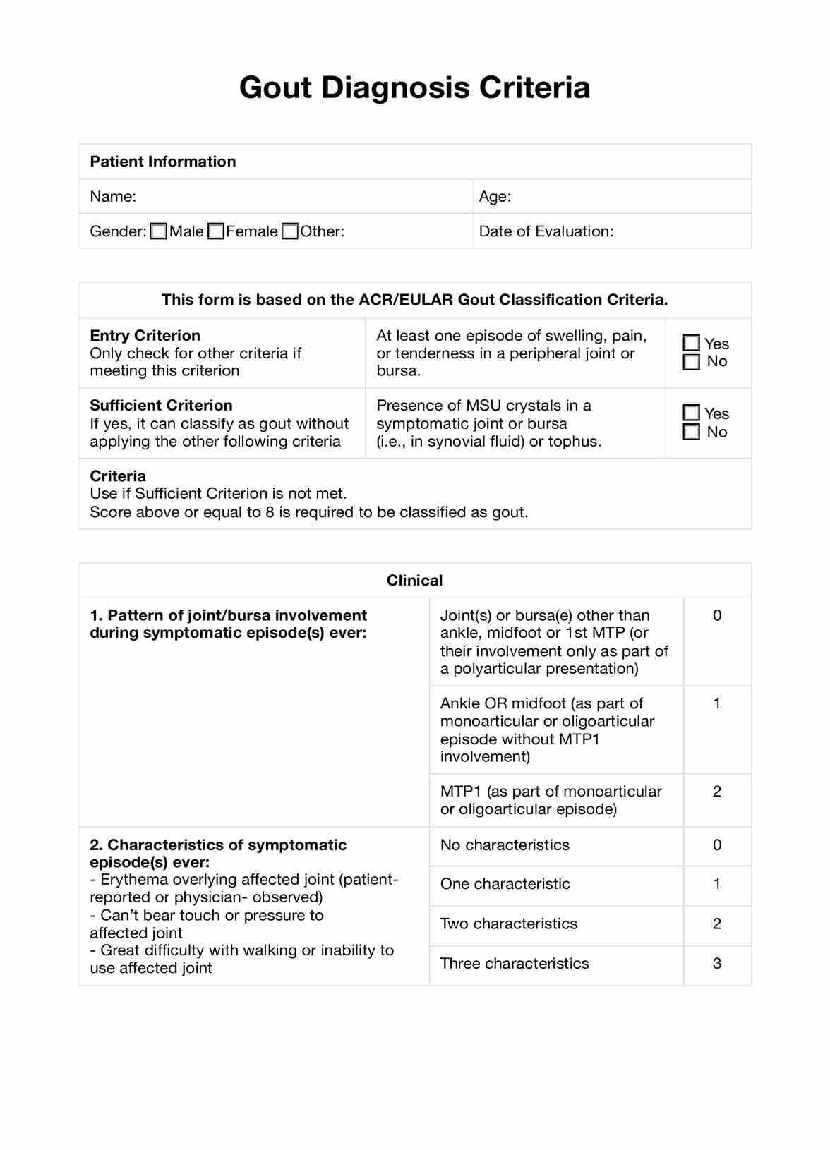
Gout diagnosis is confirmed through a combination of clinical evaluation, laboratory tests (such as serum urate levels), and imaging studies (like ultrasound or X-ray) to detect urate crystals or joint damage.

What is the most reliable diagnostic indicator of gout?

The most reliable diagnostic indicator of gout is the presence of monosodium urate (MSU) crystals in the synovial fluid of a symptomatic joint or in a tophus, as observed under a microscope.

What is the gold standard for diagnosing gout?

The gold standard for diagnosing gout is the identification of monosodium urate crystals in synovial fluid or tophaceous material using polarized light microscopy.

Join 10,000+ teams using Carepatron to be more productive

One app for all your healthcare work