## **What is a Knee Muscle Diagram?**
For some of the more complicated joints in the body, the best way to orient the various layers of muscles and tendons is by using a visual aid like this Knee Muscle Diagram.
Anatomical diagrams can provide a clear, albeit often simplified, view of key anatomical features to aid comprehension. Additionally, having an idea of the locations of the ligaments of the knee can be a great way to aid a patient's understanding of their injuries and encourage adherence to a recovery plan.
This Knee Muscle Diagram focuses on the ligaments of the knee, both lateral and cruciate ligament pairs, and the quadriceps muscle, patellar tendon, and accessory structures such as the menisci and articular cartilage.
Alternatively, this resource can also be used for practicing clinical management of various knee injuries or simply to revise the anatomy of the knee.
## **How to use our Knee Muscle Diagram template**
You can incorporate this simple knee muscle anatomy diagram into your study or practice in many ways. Here's how to get started:
### **Step 1: Download the diagram**
The first step is for you to get your hands on this anatomical Knee Muscle Diagram. To do so, simply click "Use template" to open it in the Carepatron app, where you can customize it before printing. You can also open the ready-to-print PDF version by clicking "Download."
### **Step 2: Print out or save locally**
Next, you'll want to store the diagram somewhere you can access it for your use or study. This printable knee diagram is a great addition to your practice's wall or to your lecture notes, whether digital or hard copy.
### **Step 3: Add notes, annotations, or color coding**
The last and most important step is to utilize the diagram in your studies or practice. This could look like adding extra annotations or providing this resource to your patients or students.
## **What the diagram includes**
Our Knee Muscle Diagram provides a detailed visual overview of the structures that stabilize, move, and support the knee joint, a synovial hinge joint connecting the thigh bone (femur) to the shin bone (tibia). This diagram highlights critical soft tissue components such as the quadriceps tendon, patellar tendon, and articular cartilage, which cushion and stabilize the joint during activities like walking, running, or when the knee bends under load.
Key stabilizing ligaments are clearly labeled, including the anterior cruciate ligament, posterior cruciate ligament, medial collateral ligament, and lateral collateral ligament, all of which maintain structural integrity during flexion and rotation. Intra-articular structures such as the menisci and joint capsule are also represented, helping learners understand how these elements protect the joint and regulate movement.
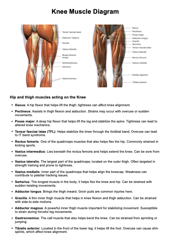
The diagram emphasizes the major muscle groups that act on the knee from the lower leg and thigh bone. These include the quadriceps group—rectus femoris, vastus lateralis, vastus medialis, and vastus intermedius—which all play critical roles in extending the knee. On the posterior side, the hamstring muscles, including the biceps femoris, assist in flexion and provide dynamic joint support.
Additional human body muscles relevant to hip and knee function are also illustrated, such as the tensor fasciae latae, sartorius, adductor longus, adductor magnus, gracilis, iliacus, pectineus, and psoas major. These muscles influence knee alignment, hip mobility, and overall posture.
Completing the view are lower limb muscles such as the gastrocnemius and tibialis anterior, which cross the knee and ankle joints, contributing to coordinated motion of the leg during gait. Together, these anatomical features provide a full picture of how the knee integrates with the rest of the musculoskeletal system for mobility and load-bearing function.
## **When are the best times to use this Knee Muscle Diagram template?**
This Knee Muscle Diagram is a versatile resource that can be used in various educational and clinical scenarios. Whether you're brushing up on anatomy, preparing for exams, or explaining conditions to patients, this diagram helps clarify complex structures of the knee joint in a visual and accessible way.
### **Lecture revision**
Getting comfortable with the basic anatomy of the knee is crucial for many university courses, such as nursing, medicine, or anatomy. This diagram can serve as a visual reference for your study, helping to provide some variety for your study resources.
### **Educational resource for patients or students**
This resource isn't just for students. Clinicians can use this diagram to educate their patients on simple knee anatomy to help them understand common injuries or conditions. This free resource can also be used by academics, lecturers, or tutors to provide to their own students.
### **Exam or test prep**
Use this diagram as preparation for an upcoming exam or test by ensuring you can name all the labeled structures of the knee or add in any extra structures, such as muscles of the lower limb or innervation of the knee.
### **Revision for orthopedics rotations**
If you're already out of nursing or med school but just need a quick refresher, this diagram can be a great go-to revision resource. It sets out the most important anatomical structures and can be annotated with any additional points you need to know before going into an orthopedics rotation.
### **Clinical reference in your healthcare practice**
This Knee Muscle Diagram can also serve as a practical reference during consultations, physical therapy, or clinical rehabilitation settings. Professionals can use it to explain anatomical structures when evaluating knee pain, planning treatment, or educating patients on injury recovery and prevention. Having a visual aid enhances patient understanding and improves communication during real-time assessments.
## **What are the benefits of using a Knee Muscle Diagram?**
Whether you're a student, clinician, or patient, visual tools like this diagram reinforce learning and support better outcomes. The Knee Muscle Diagram simplifies complex anatomy, making it easier to retain information, plan treatments, or teach others.
### **Simple design**
We have kept this resource simple but effective with two views of the knee, labeled key structures, and space for additional notes. This makes the Knee Muscle Diagram a versatile clinical or study resource.
### **Focus on basic anatomy**
Before you can move on to more advanced topics in anatomy or orthopedics, you must be completely confident in your knowledge of the knee's anatomy. This diagram includes labels for the key muscles of the knee, but can be annotated with additional labels or structures.
### **Full color**
We have used a beautiful, full-color diagram of the knee in this resource to provide a more realistic view of the knee structures and emphasize depth and the three-dimensional nature of these structures.
### **Front and side views**
In order to best facilitate a three-dimensional understanding of the ligaments of the knee, this diagram provides both front (coronal) and side (sagittal) views of the knee joint, from the lateral aspect. It also includes the muscles that interact with the knee. Ensuring you can confidently name the ligaments of the knee (as well as its surrounding muscles) is a great way to test your depth of understanding.