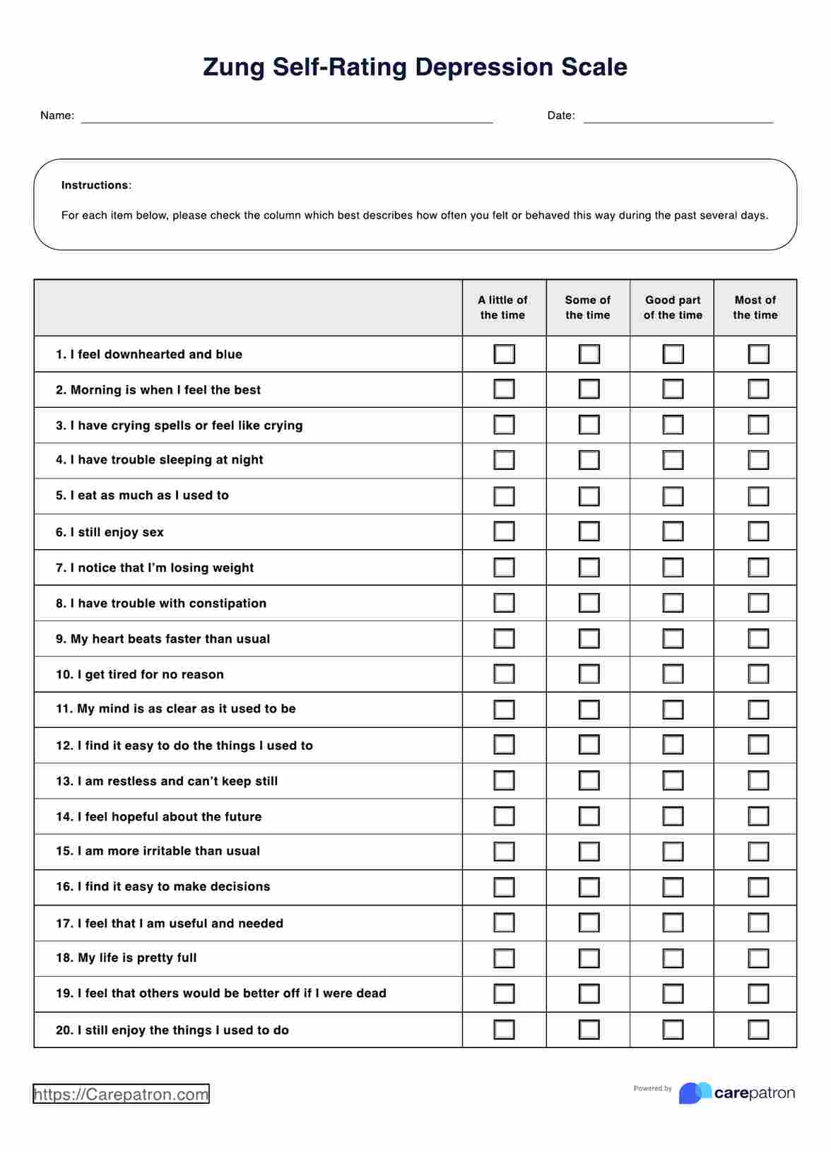
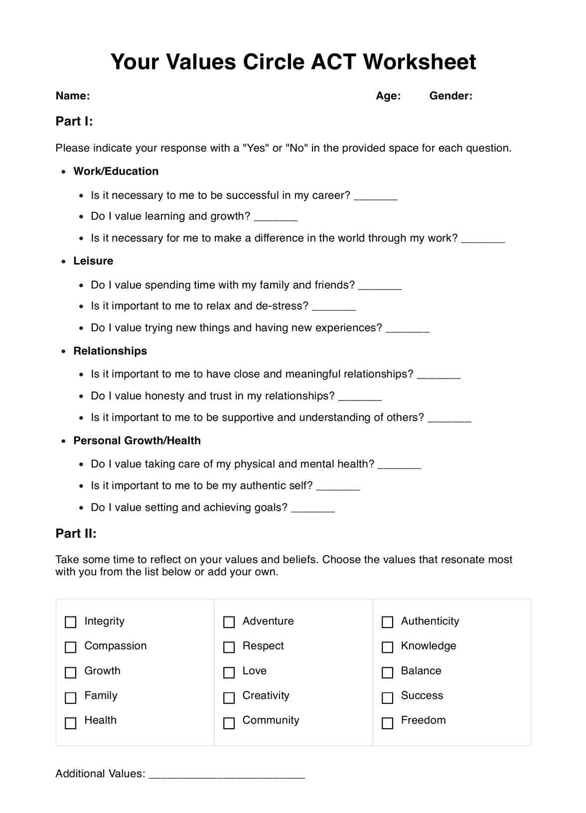
What is a Retinal Detachment Test?

Retinal detachment is most usually detected when an individual notices changes in their vision. Typically, individuals report seeing squiggly lines, flashes of light, or have trouble seeing through one eye. Although this may not always be accompanied by pain, it is essential that individuals seek urgent medical attention following any vision impairments.

A Retinal Detachment Test is a diagnostic procedure that operates to assess the condition of the eye, and determine whether there is retinal detachment. Retinal detachments occur when the retina is pulled away from the back of your eye, away from supportive tissues. Although this can manifest in different ways, this injury is most commonly a result of prolonged eye injuries, such as a retinal tear, or due to age. There has also been some evidence to suggest that individuals with a family history of retinal detachment may be genetically predisposed to this injury, however, this has ample evidence and requires further investigation.

Prolonged injury that is not assessed or treated can lead to changes in your vision, and increased risks of permanent blindness. Therefore, it is essential that an accurate Retinal Detachment Test be completed as soon as it is appropriate to do so.

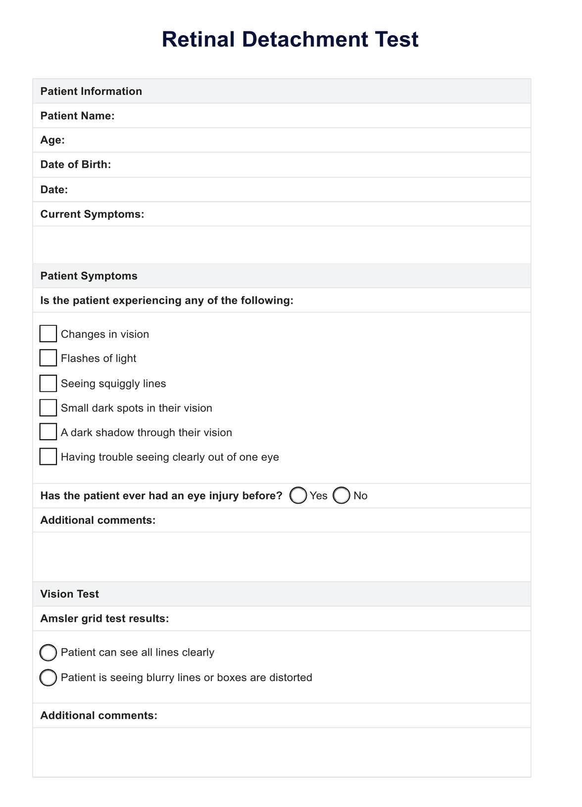
Using our Retinal Detachment Test template, health practitioners such as optometrists or photographic technicians can assess the level and type of damage to the retina. This may help diagnose retinal detachment, and prompt early treatment to support healing of the eye. Furthermore, it may also identify individuals who may be at risk of developing retinal detachment in the future, where the eye doctor may recommend proactive steps to mitigate risk factors.

Printable Retinal Detachment Test

Download this Retinal Detachment Test for health practitioners such as optometrists or photographic technicians can assess the level and type of damage to the retina.

How does this test work?

Using our Retinal Detachment Test template, health practitioners can promote early detection of retinal detachment and related eye injury. This can prompt treatment through eye surgery, eye drops, or recommendations for reading classes to mitigate risks of further injury, and help prevent permanent vision loss.

To show you how these testing procedures operate, we have broken down the process into the following steps:

Step 1: Obtain the resource

Begin by accessing the resource electronically here, or by downloading the PDF using the link below:

Download the Retinal Detachment Test PDF here.

Step 2: Patient symptoms

During your next patient consultation, if patients are experiencing any symptoms related to retinal detachment or vision loss, it is essential to complete a retinal detachment examination alongside a vision screening.

Step 3: Vision test

Complete a vision screening using the Amsler grid test. You can find an Amsler grid chart here, that can be printed and hung in your place of work.

Step 4: Dilated eye exam

Begin testing procedures by completing a dilated eye exam. This may involve dilation of the pupils by placing eye drops, allowing practitioners to view the retina to observe any tears or issues that may be causing symptoms.

Step 5: Further testing

Following the eye exam, it may be beneficial to conduct further testing to gain insight into the extent of damage and type of injury. This may involve using one or more of the following procedures:

  • Computed tomography (CT scan)
  • Eye ultrasound scan:
  • Fluorescein angiography: assess issues with blood vessels in the retina.
  • Fundus imaging:
  • Optical coherence tomography (OCT scan):

Take care to make notes of the types of tests used, and the results to update patient records.

Step 6: Diagnosis and recommendations

Following testing procedures, explain the diagnosis and what that means to your patients. In this step, it is essential to explain the mechanism of injury, and discuss recommended treatment options with patients to decide what option suits them best. This step may also involve writing referrals for specialist treatment or neurological surgery.

Step 7: Ongoing monitoring and support

Following testing, it is essential to continue monitoring patient experiences by scheduling regular eye exams, ophthalmologic exams, or consultations to support the patient.

Retinal Detachment Test example (sample)

Our free Retinal Detachment Test template is an invaluable resource that can help assist health practitioners, such as eye doctors and general practitioners, assess the eye and diagnose retinal detachment. To show how this template may operate in practice, we have created an example document.

This example uses fictional information from a patient experiencing symptoms of a detached retina, including flashes of light, squiggly lines and vision changes in one eye compared to the other eye. The patient notes that this started following eye trauma the experienced while playing sports. As a precautionary procedure, the eye doctor has recommended a retinal detachment test alongside a regular eye exam.

It is recommended that this example be viewed prior to conducting testing, however, please consider that information in the example is entirely fictional, and should not supplement tailored health advice or procedures. You can view the example here, or download the resource using the following link:

Download our free Retinal Detachment Test example here

Retinal Detachment Test example (sample)

Results and next steps

The results that emerge from a Retinal Detachment Test are multi-faceted, and can be experienced differently between patients according to various individual factors. These may include the patient's family history, stage of eye injury, and cause of the injury. However, the following are some results that may be observed, and what they may mean for the individual:

Retinal tear

A retinal tear occurs when the vitreous peels away from the retina, where the force of this pulling motion can create a tear through the retina. If diagnosed with a retinal tear, it is essential that this be treated as soon as possible, as the tear allows liquid vitreous to pass through and can lead to retinal detachment.

In this instance, an eye doctor may recommend laser surgery to connect the retina to underlying tissue, or use a freezing probe over the eye surface to connect the retina to the wall of the eye. This will support recovery and prevent retinal detachment.

Retinal detachment

If diagnosed with retinal detachment, individuals will require early treatment through surgery. Depending on the severity of the detached retina, as well as other factors such as patient history, the type of surgery recommended may differ.

The following surgeries may be recommended:

  • Pneumatic retinopexy: The surgeon sutures silicon to the sclera (white of your eye), which indents the eye wall, reducing the pull on the retina. If there is a large detachment or several damages, the surgeon may choose to leave a permanent scleral buckle to hold the entire eye.
  • Scleral buckle surgery: The procedure involves the vitreous cavity of the eye being injected with air or gas, causing the retina to press against the eye wall. This stops the vitreous liquid, and allows the surgeon to repair the tear.
  • Vitrectomy: This process involves the removal of any tissue and vitreous that are pulling at the retina, and then injecting air, gas, or silicone oil into the vitreous area.

Lattice degeneration

Patients may be experiencing lattice degeneration, or retinal thinning, which can lead to retinal tears or detachment. To prevent further damage, the eye doctor may schedule annual appointments to check the status of the eye, or conduct a dilated eye exam to further assess the situation before discussing treatment options.

Macular degeneration

Patients with macular degeneration may be experiencing damage to the macula within the retina. While there are no direct treatments for macular degeneration, there are procedures that may help slow down vision loss and progression of the condition.

Preventing retinal detachment

Ageing is often the main cause of retinal detachment, which can make it difficult to prevent. However, there are a few things you can do to help reduce risks of retinal detachment, and slow the progression of vision loss. These may include:

  • Scheduling regular eye exams with the optometrist: This can promote early identification and treatment of eye diseases or related conditions. Often conditions like myopia or retinal tears can make individuals more susceptible to retinal detachment, thus early detection can allow for treatment early.
  • Safety wear: When participating in risk activities such as laboratory work, painting, or in high-dust environments, it is essential to wear safety goggles. These can protect your eyes from hazardous particles.
  • Immediate treatment: If there are any changes in vision, it is critical to get treatment as soon as you can. Whether this means scheduling an appointment with the optometrist, general practitioner, or going to the emergency room, early treatment has a critical role in mitigating risks.
What is a Retinal Detachment Test?

Commonly asked questions

What is a Retinal Detachment Test?

A Retinal Detachment Test is an eye examination procedure used to identify whether there is damage at the site of the retina, specifically if the retina has become detached from the blood vessels.
Who conducts the test?

This is most typically conducted by an eye doctor following symptoms of vision changes.

When should I have a Retinal Detachment Test?

These tests are usually recommended upon referral, or conducted by your eye doctor following any changes in vision. This includes any obstructions to vision, seeing dots or squiggly lines, or blurred sight.

Join 10,000+ teams using Carepatron to be more productive

One app for all your healthcare work