


What is a pulmonary function test (PFT)?
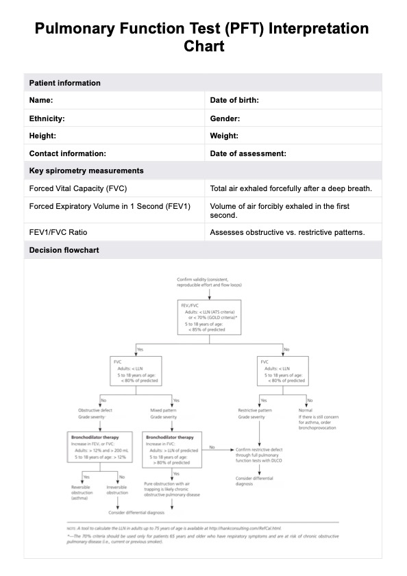
Pulmonary function tests (PFTs) are specialized lung function tests that measure how well the lungs perform critical functions such as air movement, gas exchange, and volume changes during respiration (Stanojevic et. al., 2022). This pulmonary function testing helps healthcare professionals assess the presence and severity of respiratory disease, including obstructive lung disease (e.g., chronic obstructive pulmonary disease) and restrictive lung disease. Commonly measured parameters include forced vital capacity (FVC), forced expiratory volume in one second (FEV1), and total lung capacity (TLC), which provide valuable insights into respiratory mechanics.
PFTs are instrumental in identifying conditions such as upper airway obstruction or early signs of pulmonary impairment due to occupational exposures or pulmonary toxicity. While they do not offer a definitive diagnosis, they guide physicians in correlating total lung capacity results with the patient’s medical history, physical examination, and additional diagnostic data.
PFTs also aid in monitoring disease progression and treatment efficacy, quantifying changes in lung function over time. Proper technique and patient effort are crucial to obtaining reliable data, as poor effort can affect test accuracy. Updated spirometry guidelines from organizations such as the European Respiratory Society and American Thoracic Society ensure standardized measurements and interpretation for consistent clinical use (Graham et. al., 2019).


