Dermatomyositis Diagnosis Criteria

Learn about the diagnostic criteria for dermatomyositis and see an example with Carepatron's free PDF download. Get the information you need to understand this condition.

By Wynona Jugueta on Apr 11, 2024.

Fact Checked by Ericka Pingol.

Use Template

What is dermatomyositis?

Dermatomyositis is a rare autoimmune disorder characterized by muscle weakness and inflammation, often accompanied by skin rash. It falls under the umbrella of idiopathic inflammatory myopathies, a group of muscle diseases of unknown origin. This condition primarily affects the skeletal muscles, causing progressive muscle weakness and fatigue.

One of the distinguishing features of dermatomyositis is its unique skin manifestations, which include a distinctive rash on the face, eyelids, knuckles, elbows, knees, and other areas exposed to the sun.

Muscle involvement in dermatomyositis can range from mild weakness to severe debilitation, impacting daily activities and quality of life. Diagnosis typically involves a combination of clinical evaluation, laboratory tests, imaging studies, and sometimes a muscle biopsy to confirm inflammation. Elevated levels of muscle enzymes such as creatine kinase (CK) and aldolase in the blood are often observed in dermatomyositis, indicating muscle damage.

In addition to muscle symptoms, dermatomyositis may also affect other organs, including the lungs. Interstitial lung disease is a common complication, leading to breathing difficulties and decreased lung function. Early detection and management of these complications are essential for optimal outcomes.

Physical therapy plays a crucial role in maintaining muscle strength and function, improving mobility, and enhancing overall quality of life for individuals with dermatomyositis. It helps to preserve muscle mass, prevent contractures, and alleviate pain.

Dermatomyositis symptoms

Dermatomyositis symptoms can vary widely among individuals and may involve multiple body systems. Recognizing these symptoms is crucial for early diagnosis and effective management of the condition.

Here, we outline the key symptoms associated with dermatomyositis.

  • Muscle weakness: Progressive muscle weakness, especially in the proximal muscles (muscles closest to the trunk), making it difficult to perform everyday tasks such as climbing stairs, getting up from a seated position, or lifting objects.
  • Skin rash: Characteristic rashes may be red or purple, often accompanied by swelling and itching.
  • Muscle pain: Pain and tenderness in muscles, particularly during movement or pressure on the affected muscles.
  • Difficulty swallowing: Dysphagia or difficulty swallowing due to muscle weakness in the throat and esophagus.
  • Fatigue: Persistent fatigue and weakness, which may not improve with rest.
  • Joint pain: Joint pain and stiffness resembling symptoms of arthritis.
  • Shortness of breath: Interstitial lung disease, leading to shortness of breath, cough, and decreased exercise tolerance.
  • Fever: Low-grade fever, especially during periods of disease activity.
  • Weight loss: Unintentional weight loss, often associated with decreased appetite and muscle wasting.
  • Gottron's papules: Raised, scaly, reddish-purple patches over the knuckles, elbows, or knees, known as Gottron's papules.
  • Heliotrope rash: Bluish-purple discoloration or swelling of the eyelids, known as a heliotrope rash.
  • Mechanic's hands: Thickened, cracked, and fissured skin on the palms and fingers, resembling the hands of a mechanic.

Risk factors and causes of dermatomyositis

Understanding the risk factors and causes of dermatomyositis is essential for identifying individuals who may be at higher risk and for developing strategies for prevention and early intervention.

Below are some factors that may contribute to the development of dermatomyositis:

  • Autoimmune factors: Dermatomyositis is believed to result from an abnormal immune response, where the body's immune system mistakenly attacks its own muscle and skin tissues.
  • Genetic predisposition: While dermatomyositis can occur in individuals without a family history, there may be genetic factors that predispose certain individuals to develop the condition.
  • Environmental triggers: Exposure to certain environmental factors, such as ultraviolet (UV) radiation from sunlight, infections, or medications, may trigger or exacerbate dermatomyositis in susceptible individuals.
  • Age and gender: Although dermatomyositis can occur at any age, it most commonly affects adults between the ages of 40 and 60, as well as children between the ages of 5 and 15. Additionally, women are more commonly affected than men.
  • Underlying conditions: Dermatomyositis may be associated with other autoimmune diseases, such as systemic lupus erythematosus, or with certain types of cancer, including ovarian cancer. Individuals with these conditions may have an increased risk of developing dermatomyositis.
  • Muscle enzyme abnormalities: Elevated levels of muscle enzymes, such as creatine kinase (CK) and aldolase, in the blood may indicate muscle damage and inflammation, which are characteristic features of dermatomyositis.
  • Symmetrical muscle weakness: The presence of this muscle weakness, particularly affecting the proximal muscles (muscles closest to the trunk), is a hallmark feature of dermatomyositis.

Complications it can lead to

While dermatomyositis primarily affects the muscles and skin, it can lead to various complications that affect multiple body systems. Understanding these potential complications is crucial for comprehensive management and improving outcomes for individuals with dermatomyositis.

Here are some of the complications associated with this condition:

  • Interstitial lung disease (ILD): Dermatomyositis can lead to ILD, a group of lung disorders characterized by inflammation and scarring of the lung tissue. ILD can cause breathing difficulties, coughing, and decreased lung function, impacting respiratory health.
  • Respiratory muscle weakness: In severe cases, progressive weakness of the respiratory muscles, including the diaphragm and intercostal muscles, can lead to respiratory failure and the need for mechanical ventilation.
  • Aspiration pneumonia: Dysphagia (difficulty swallowing) due to muscle weakness can increase the risk of aspiration pneumonia, where food, liquids, or saliva are inhaled into the lungs, leading to lung infections.
  • Dysphonia: The weakness of speech production muscles can cause dysphonia (hoarseness) or difficulty speaking clearly.
  • Dysphagia and malnutrition: Severe dysphagia may lead to inadequate food intake, malnutrition, and weight loss, affecting overall health and well-being.
  • Contractures: Prolonged muscle weakness and immobility can result in contractures, where the muscles and tendons become permanently shortened, leading to joint stiffness and limited range of motion.
  • Ulcers and skin infections: Chronic skin involvement in dermatomyositis, characterized by skin manifestations such as ulcers, rashes, and sores, can increase the risk of skin infections and delay wound healing.
  • Calcinosis: In some cases, calcium deposits may form in the skin and soft tissues (calcinosis), leading to pain, inflammation, and impaired mobility.
  • Malignancy: Dermatomyositis has been associated with an increased risk of certain cancers, particularly ovarian and lung cancer. Regular screening for cancer may be recommended in individuals with dermatomyositis.

Printable Dermatomyositis Diagnosis Criteria

Download this Dermatomyositis Diagnosis Criteria to enhance your diagnostic accuracy and ensure timely intervention for patients presenting with suspected dermatomyositis.

What tests can help diagnose dermatomyositis?

Dermatomyositis can present with a variety of symptoms, making diagnosis challenging. Healthcare providers may employ various tests to confirm the presence of the disease and rule out other conditions. Here are some common tests used in the diagnosis of dermatomyositis:

Muscle enzyme levels tests

Elevated levels of CK, an enzyme released into the bloodstream when muscle fibers are damaged, can indicate muscle disease or injury. In dermatomyositis, CK levels are often elevated, reflecting muscle involvement.

Electromyography (EMG)

EMG measures the electrical activity of muscles. In dermatomyositis, EMG may reveal abnormal electrical patterns characteristic of muscle inflammation and weakness.

Muscle biopsy

Muscle biopsy findings involve removing a small sample of muscle tissue for examination under a microscope. In dermatomyositis, muscle biopsy can reveal characteristic features such as muscle fiber necrosis, inflammation, and fibrosis, confirming the diagnosis and distinguishing it from other muscle diseases.

Skin biopsy

A skin biopsy involves taking a small sample of skin tissue for examination under a microscope. In dermatomyositis, skin biopsy may reveal characteristic changes such as interface dermatitis, basement membrane thickening, and perivascular inflammation, aiding in diagnosing the condition.

Blood tests

Blood tests are essential in diagnosing dermatomyositis as they can provide valuable insights into the immune response and autoantibody presence associated with the condition. The antinuclear antibody (ANA) test, which detects antibodies targeting the body's cells, is commonly utilized, as elevated levels may indicate autoimmune activity. Moreover, autoantibody testing, including specific antibodies such as anti-Jo-1 antibodies, can aid in confirming dermatomyositis and distinguishing it from other muscle diseases.

Imaging studies

Imaging studies, such as MRI, X-rays, and CT scans, play a crucial role in assessing the extent of inflammation the muscle and identifying potential complications of dermatomyositis. MRI is beneficial for detecting idiopathic inflammatory myopathy and evaluating the severity of muscle involvement. At the same time, X-rays and CT scans can help assess lung involvement and detect underlying dermatomyositis-associated malignancies.

Physical examination

A thorough physical examination is paramount in diagnosing clinically amyopathic dermatomyositis, as it allows healthcare providers to assess characteristic signs such as proximal muscle weakness and skin disease. Muscle weakness, especially around the hips and shoulders, is a hallmark of dermatomyositis and is often evaluated through manual muscle testing and functional assessments.

What is the diagnostic criteria for dermatomyositis?

The criteria for dermatomyositis involve a combination of clinical, laboratory, and imaging findings to accurately confirm the presence of the disease. The requirements typically include the following:

Characteristic skin manifestations

Dermatomyositis is often characterized by distinctive skin changes, such as the heliotrope rash (a purplish discoloration of the eyelids), Gottron's papules (raised, scaly patches on the knuckles), and a characteristic rash on the face, neck, shoulders, and upper chest.

Proximal muscle weakness

Symmetrical muscle weakness affecting the proximal muscles (closest to the trunk) is a hallmark feature of dermatomyositis. This weakness may manifest as difficulty in rising from a chair, climbing stairs, or lifting objects.

Muscle biopsies

Muscle biopsies may reveal characteristic histopathological findings, including muscle fiber necrosis, inflammation, and perivascular infiltration of inflammatory cells, supporting the diagnosis of dermatomyositis.

Laboratory tests

Elevated levels of muscle enzymes, such as creatine kinase (CK) and aldolase, in the blood are often observed in dermatomyositis, reflecting muscle inflammation and damage.

Imaging studies

Imaging modalities, such as magnetic resonance imaging (MRI) and electromyography (EMG), may help assess muscle involvement and confirm the diagnosis.

Screening for underlying malignancy

Given the association between dermatomyositis and certain malignancies, such as ovarian cancer and lung cancer, screening for underlying malignancy may be warranted in patients with dermatomyositis, especially in older individuals.

Dermatomyositis Diagnosis Criteria example

To illustrate how these criteria can be applied in a clinical setting, we've created a sample Dermatomyositis Diagnosis Criteria template at Carepatron. This template is a practical tool for healthcare practitioners, guiding them through the key steps and considerations in diagnosing dermatomyositis.

Our Dermatomyositis Diagnosis Criteria template integrates the essential components of criteria, including clinical features, laboratory tests, and imaging studies. By following this structured approach, healthcare providers can systematically evaluate patients presenting with suspected dermatomyositis, ensuring thorough assessment and accurate diagnosis.

This template not only aids in the diagnostic process but also promotes consistency and standardization in clinical practice, enhancing efficiency and reducing the risk of oversight or misinterpretation.

Download our free Dermatomyositis Diagnosis Criteria template example here

Dermatomyositis Diagnosis Criteria example

Dermatomyositis treatments

Dermatomyositis, a complex autoimmune disorder, necessitates a multifaceted treatment approach to manage both its skin and muscle manifestations alongside systemic symptoms. Here are some common treatments:

Medications

Corticosteroids such as prednisone are typically the first-line treatment, reducing inflammation and suppressing the immune response. If corticosteroids are insufficient or cause significant side effects, immunosuppressive medications like methotrexate or mycophenolate mofetil may be prescribed. Biological therapies targeting specific immune pathways, like rituximab or intravenous immunoglobulin (IVIG), may be used in refractory cases.

Physical therapy

Physical therapy is essential for managing dermatomyositis by improving muscle strength, flexibility, and function. Therapists tailor exercise programs to target specific muscle groups affected by the disease, aiding overall mobility and quality of life.

Topical treatments

Topical corticosteroids or sun protection measures like protective clothing and broad-spectrum sunscreen alleviate skin lesions and minimize inflammation that sunlight exacerbates.

Systemic symptom management

Analgesic medications help manage muscle pain and discomfort, while targeted treatments address systemic symptoms like interstitial lung disease or dysphagia.

Monitoring and supportive care

Regular monitoring by healthcare providers ensures treatment efficacy and adjustment as necessary. Additionally, supportive care measures, including nutritional support, psychological assistance, and patient education, contribute to overall patient well-being and quality of life.

How do you diagnose dermatomyositis?

Commonly asked questions

How do you diagnose dermatomyositis?

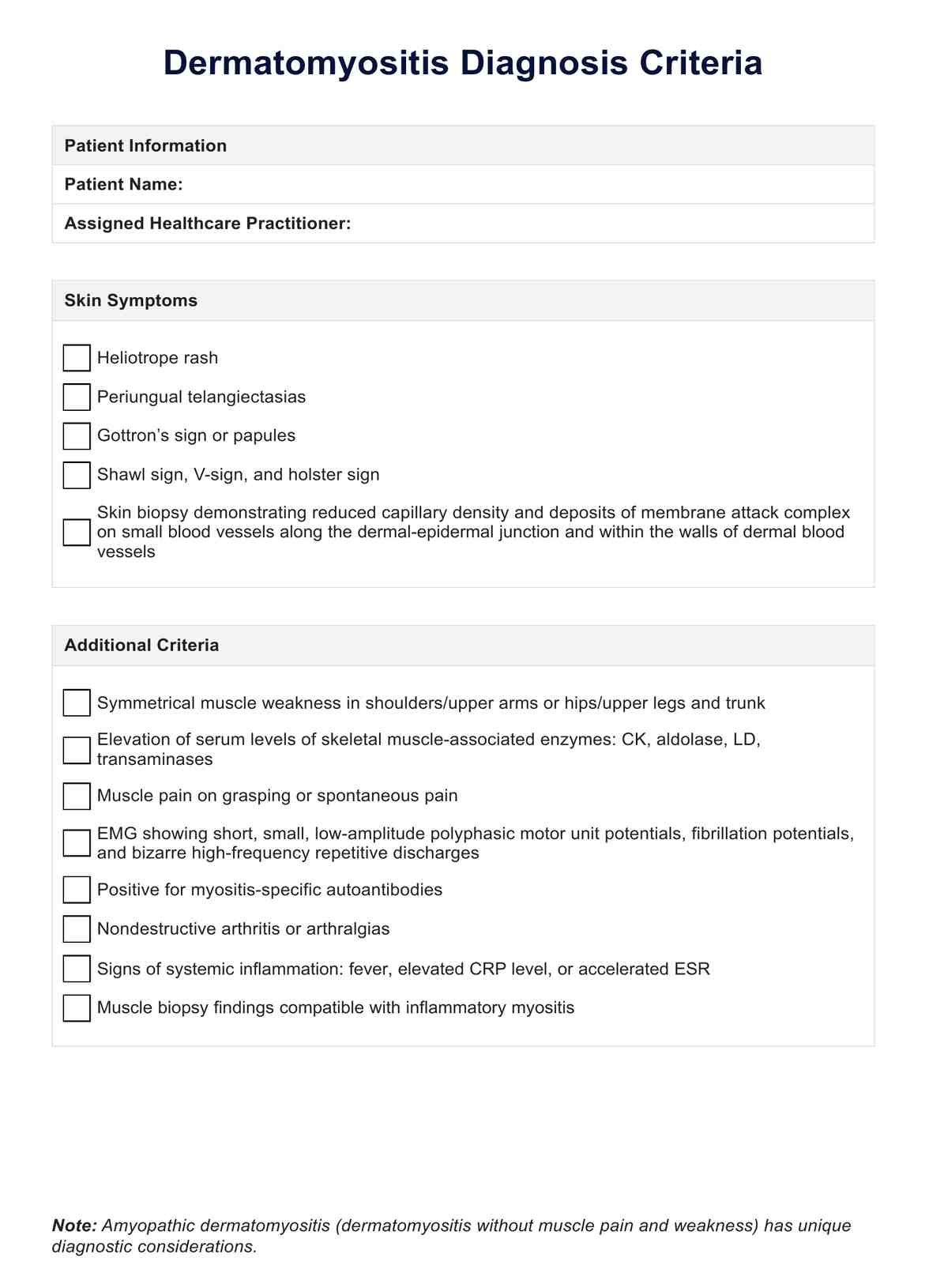
Dermatomyositis is diagnosed through clinical evaluation, muscle and skin biopsies, imaging studies, and laboratory tests.

What is the gold standard for diagnosing dermatomyositis?

A muscle biopsy is the gold standard for diagnosing dermatomyositis, which can reveal characteristic histopathological findings such as muscle inflammation and necrosis.

What labs are abnormal with dermatomyositis?

Common abnormal laboratory findings in dermatomyositis include elevated levels of muscle enzymes such as creatine kinase (CK) and aldolase and abnormal electromyography (EMG) results.

Join 10,000+ teams using Carepatron to be more productive

One app for all your healthcare work