Pleural Fluid Analysis

Unlock diagnostic insights with pleural fluid analysis. Comprehensive and timely results for effective management of respiratory conditions.

By Joshua Napilay on Apr 08, 2024.

Fact Checked by Ericka Pingol.

Use Template

What is a Pleural Fluid Analysis?

Pleural Fluid Analysis is a diagnostic procedure conducted to assess the composition of fluid that accumulates in the pleural cavity, a thin space between the two layers of the pleura surrounding the lungs. 

The pleura is a membrane that facilitates smooth lung movement within the chest cavity. Under normal circumstances, a small amount of fluid is present in this space to allow frictionless movement during breathing. However, various medical conditions can lead to an abnormal accumulation of pleural fluid, a condition known as pleural effusion.

Physicians must determine the underlying cause of pleural effusion. The procedure involves extracting a pleural fluid sample through a minimally invasive technique called thoracentesis. Once obtained, the fluid undergoes thorough examination, focusing on its physical characteristics, chemical composition, and microscopic appearance.

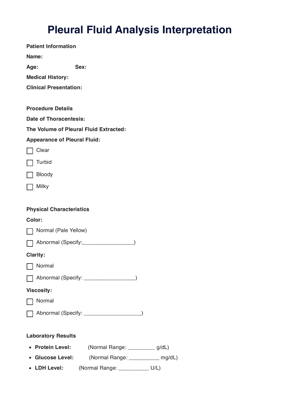
The analysis includes assessing the fluid's color, clarity, and viscosity. Additionally, laboratory tests are performed to measure the proteins, glucose, lactate dehydrogenase (LDH) levels, and cell types in the fluid. The differential cell count helps identify abnormal cells, such as those indicative of infection, inflammation, or malignancy.

It is crucial in diagnosing various conditions, including infections (such as pneumonia or tuberculosis), inflammatory diseases (like rheumatoid arthritis), heart failure, and multiple cancers. The results guide healthcare professionals in tailoring appropriate treatment plans based on the specific underlying cause of the pleural effusion.

Printable Pleural Fluid Analysis PDF

Enhance your care with our free Pleural Fluid Analysis PDF

How is a Pleural Fluid Analysis conducted?

A Pleural Fluid Analysis is a diagnostic procedure crucial for identifying the underlying causes of pleural effusion, involving the extraction and examination of fluid from the pleural cavity through thoracentesis.

Procedure

  • Patient positioning and preparation: Before the procedure, the patient is positioned upright or on their side. The targeted area is cleansed, and a local anesthetic is administered to minimize discomfort.
  • Needle insertion: A thin, hollow needle is precisely inserted through the chest wall into the pleural space using ultrasound guidance. This allows for the controlled extraction of pleural fluid.
  • Fluid collection: The needle is connected to a syringe or vacuum bottle, facilitating the withdrawal of a pleural fluid sample. The amount extracted depends on the clinical context but typically ranges from a few milliliters to several hundred milliliters.

Analysis

  • Physical characteristics: The extracted fluid is analyzed for physical attributes such as color, clarity, and viscosity, providing initial insights into its nature.
  • Laboratory tests: Laboratory analyses include measuring protein, glucose, and lactate dehydrogenase (LDH) levels in the pleural fluid. These tests contribute to the diagnostic process by indicating potential causes, such as infection, inflammation, or malignancy.
  • Microscopic examination: A microscopic examination identifies and quantifies different cell types in the pleural fluid. Abnormal cells may suggest specific underlying conditions, aiding in diagnosis.

Diagnostic significance

The comprehensive results of the analysis enable healthcare professionals, such as pulmonologists or interventional radiologists, to tailor appropriate treatment plans based on the specific cause of pleural effusion. This targeted approach ensures effective medical intervention.

How are the results of a Pleural Fluid Analysis interpreted?

Interpreting the results of an analysis is a critical step for healthcare professionals in identifying the underlying cause of pleural effusion. The interpretation involves a comprehensive analysis of various parameters obtained from the extracted pleural fluid.

Normal values

Healthcare professionals compare the results to established normal values for pleural fluid constituents. Normal pleural fluid is typically clear, pale yellow and has specific ranges for protein, glucose, and LDH levels. A normal differential cell count reveals a minimal number of cells, predominantly lymphocytes.

Abnormal pleural fluid analysis results

  • Infection: Elevated white blood cell counts and neutrophil predominance may indicate bacterial or fungal infections, while an increased lymphocyte count could suggest tuberculosis or viral infections.
  • Inflammation: Inflammatory conditions such as rheumatoid arthritis or lupus may be suggested by an elevated protein concentration and increased white blood cells.
  • Malignancy: Malignant cells in the pleural fluid indicate a potential malignancy from the lungs, breast, or other adjacent structures.
  • Heart failure: Elevated protein levels and high LDH may be indicative of heart failure-related pleural effusion.
  • Chylothorax or hemothorax: Specific characteristics such as milky appearance (chylothorax) or bloody fluid (hemothorax) provide crucial diagnostic clues.

To formulate a precise diagnosis, healthcare professionals consider these findings in conjunction with the patient's medical history, clinical presentation, and other diagnostic tests. 

Interpreting the results of the analysis is integral to tailoring appropriate treatment strategies and ensuring timely and effective management of the underlying condition contributing to pleural effusion. 

Regular communication between healthcare providers and patients is essential for a comprehensive understanding of the results and the subsequent course of medical intervention.

Pleural Fluid Analysis example (sample)

In this case, Mr. John Doe, a 55-year-old male with respiratory symptoms, underwent a Pleural Fluid Analysis on February 15. Fluid extraction revealed elevated protein (4.2 g/dL) and LDH (400 U/L) levels, with notable neutrophil and lymphocyte predominance. Microscopic examination detected suspicious atypical cells, suggesting a potential bacterial infection or underlying malignancy. 

The physician recommended further investigations, including a CT scan and sputum culture, and initiated broad-spectrum antibiotics. Follow-ups for bronchoscopy and biopsy were scheduled to assess infection or malignancy. This case underscores the diagnostic complexity of pleural effusion and the pivotal role of the analysis in guiding subsequent investigations and treatment decisions.

Download our free Pleural Fluid Analysis example PDF

Pleural Fluid Analysis example

When are the best times to conduct a Pleural Fluid Analysis?

Pleural Fluid Analysis is most commonly indicated when a patient presents with signs and symptoms suggestive of pleural effusion. Upon physical examination, these include dyspnea, chest pain, persistent cough, and diminished breath sounds. When these clinical manifestations are observed, an analysis becomes crucial to determine the underlying cause of the effusion.

Unexplained respiratory symptoms

An analysis is essential in cases where a patient experiences unexplained respiratory symptoms, such as prolonged coughing or difficulty breathing, and initial assessments, such as chest X-rays, indicate the presence of pleural effusion. This diagnostic procedure helps differentiate between various causes, such as infections, inflammatory conditions, or malignancies, guiding healthcare professionals toward targeted and effective interventions.

Monitoring known conditions

For individuals with pre-existing conditions known to be associated with pleural effusion, such as congestive heart failure or certain cancers, regular monitoring through the analysis is recommended. This proactive approach enables healthcare providers to detect changes in the pleural fluid composition early, facilitating timely adjustments to the treatment plan and ensuring optimal patient care.

Post-therapeutic assessment

After initiating treatment for conditions causing pleural effusion, healthcare professionals may analyze to assess the therapeutic response. Monitoring changes in fluid characteristics and cell counts can provide valuable insights into the effectiveness of the treatment and guide adjustments if needed.

The best times to conduct an analysis include diagnosing pleural effusion, investigating unexplained respiratory symptoms, monitoring known associated conditions, and assessing therapeutic responses. Timely and strategic use of this diagnostic tool enhances the accuracy of diagnoses and improves patient outcomes.

Why use Carepatron as your blood test app?

Experience unparalleled efficiency and precision in blood test management with Carepatron. Our specialized software and apps, including CEA and Albumin Blood Tests, offer a seamless experience for healthcare professionals and patients. 

By providing a structured framework for documenting patients' clinical history and test results, we enhance accuracy while promoting secure and convenient access to blood test data, fostering improved communication and patient engagement throughout the healthcare journey.

We offer a suite of tools for healthcare professionals beyond blood test management. Our tools include appointment scheduling, clinical note templates, secure storage, and advanced medical billing and coding features. Our interface is user-friendly and adaptable to unique requirements while adhering to guidelines for safeguarding patient information.

Unlock the potential for clinical documentation improvement with our Cloud-based EHR system. Store all medical intake forms, clinical assessments, and treatment plans securely in one place, providing healthcare providers with accurate and up-to-date information for better patient outcomes. 

Foster collaboration across healthcare teams, professional networks, and even with patients' loved ones, enhancing communication and elevating the standard of patient care. Rest easy knowing that our electronic health records software is HIPAA-compliant, ensuring the safe and secure storage of patient information.

Choose Carepatron for a comprehensive, user-friendly, and secure platform that empowers healthcare providers to deliver superior patient care.

Clinical notes software benefit
Why are Pleural Fluid Analyses important?

Commonly asked questions

Why are Pleural Fluid Analyses important?

Pleural Fluid Analyses are crucial for diagnosing the underlying causes of pleural effusion and guiding targeted treatment plans for conditions like infections, inflammation, and malignancies.
How long does it take to get Pleural Fluid Analysis results?

The results typically take a few days, but urgency may expedite the process. Timely communication is maintained to address patient needs.

What needs to be done to prepare for a Pleural Fluid Analysis?

Minimal preparation is required. Patients may be asked to refrain from eating or drinking for a few hours before the procedure. Informing the healthcare provider about medications is essential.

Are there risks to taking/conducting a Pleural Fluid Analysis?

While generally safe, risks include infection, bleeding, or a temporary increase in respiratory distress. These risks are minimal and outweighed by the diagnostic benefits.

Join 10,000+ teams using Carepatron to be more productive

One app for all your healthcare work