What are syndesmosis injuries?
Syndesmosis injuries, also known as high ankle sprain, affect the distal tibiofibular ligaments and typically arise from sudden external rotation of the ankle. These injuries are not as common as typical ankle sprains but can cause significant discomfort and impairment. External rotation forces during injury cause the talus to rotate laterally, pushing the fibula away from the tibia. This can lead to increased stresses on the tibia, likelihood of lateral subluxation of the distal fibula, and incongruence of the ankle joint articulation.
Symptoms of syndesmosis injuries include anterolateral ankle pain proximal to the anterior inferior tibiofibular ligament, accompanied by tenderness and swelling. Patients may struggle to bear weight, particularly when the interosseous membrane is involved. Diagnosis relies on clinical suspicion, palpation for tenderness over the syndesmosis, and specific provocative tests like the squeeze and external rotation stress tests.
Imaging, such as radiographs and MRIs, aids in confirming the diagnosis, especially when syndesmotic injuries are suspected. Treatment varies depending on the severity of the injury. Nonoperative approaches involve immobilization with a CAM boot or cast, and physical therapy programs. You may also resort to operative management, including syndesmosis screw fixation or suture button fixation, for more severe cases with instability or associated fractures.
How to diagnose syndesmosis injuries
Diagnosing syndesmosis injuries involves a comprehensive assessment, including history, observation, palpation, and special testing. Clinical history, particularly focusing on mechanisms of injury, such as external rotation, is crucial. Observation may reveal less swelling than lateral ankle sprains and limitations in plantar flexion and weight-bearing ability. Palpation over specific areas, including the anterior tibiofibular ligament and the interosseous membrane, aids in identifying tenderness.
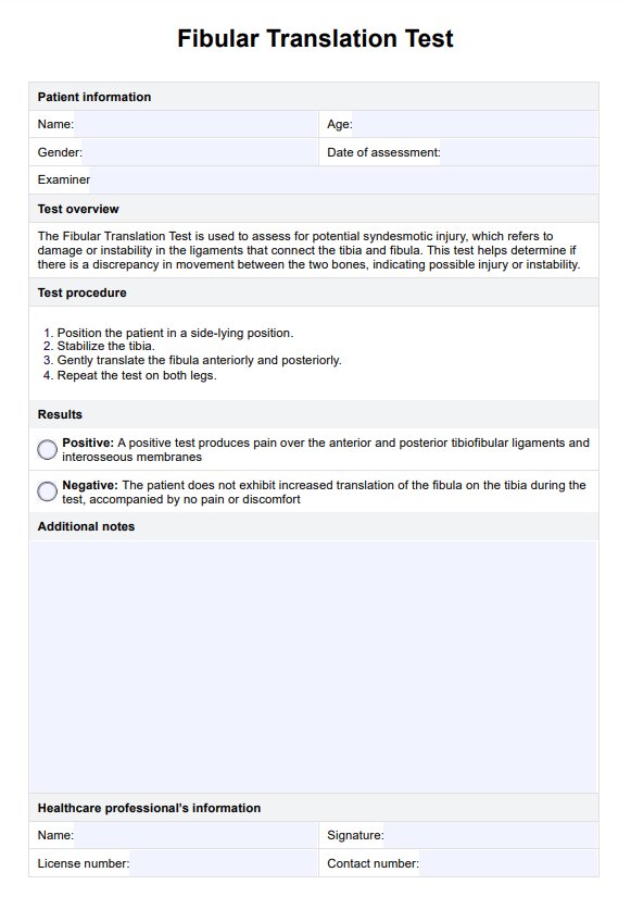
Special tests like the Fibular Translation Test assess for increased translation, indicative of syndesmosis instability. Imaging, including plain films and MRI, confirms the diagnosis and guides treatment decisions (Williams et al., 2007).


