Diplopia Test

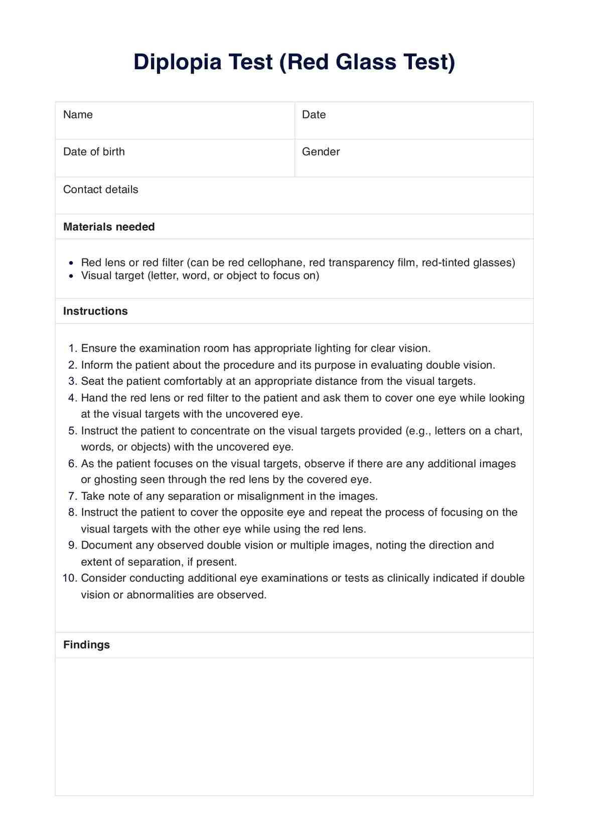
Learn how to conduct the Red Glass or Diplopia Test. Download a free PDF template and example in this guide.

By Ericka Pingol on Apr 08, 2024.

Fact Checked by Ericka Pingol.

Use Template

What is diplopia?

Diplopia, commonly known as double vision, is when a person sees two images of the same object instead of one. This can occur in one or both eyes and can be present at all times or only when looking at particular objects or in specific directions.

Diplopia, also known as double vision, is a condition where a person sees two images of a single object instead of one. This can occur in one or both eyes and may be constant or intermittent.

There are several potential causes of diplopia, each relating to different areas of the vision system (Cleveland Clinic, n.d.). It could be due to the misalignment of one or both eyes, an issue called strabismus. Problems with the muscles that control the movement of the eyes or the nerves that stimulate those muscles can also be the source of double vision.

In some cases, it may result from a systemic condition like diabetes or hypertension. Diseases such as Myasthenia Gravis, Graves' Disease, and Guillain-Barre Syndrome are also linked with diplopia.

Diplopia can also occur as a side effect of certain medications, such as anti-seizure drugs or muscle relaxants. Additionally, head injuries or neurological conditions like multiple sclerosis, thyroid eye disease, or brain tumors can lead to double vision.

Printable Diplopia Test

Download this Diplopia Test, which helps diagnose diplopia.

Monocular vs binocular diplopia

Diplopia can be classified into monocular and binocular (Cleveland Clinic, n.d.).

Monocular double vision is when the double vision occurs in one eye only, while binocular diplopia is when both eyes are affected. Issues with the cornea or lens of the affected eye, such as corneal scarring or cataracts, often cause monocular diplopia.

In contrast, binocular double vision is usually due to problems with the eye muscles or nerves controlling them. These issues can lead to ocular misalignment, causing the eyes to point in different directions and resulting in two images (Najem & Margolin, 2023).

To diagnose diplopia, a comprehensive eye examination is required. This includes a physical exam and tests like the red glass or Diplopia Test, which helps identify the affected eye.

Common causes

As mentioned,  diplopia can be a symptom of various underlying conditions. Here are some of the most common causes of diplopia in detail:

Myasthenia Gravis

An autoimmune disorder that affects the neuromuscular junction, causing muscle weakness (WebMD, n.d.). This condition often affects the extraocular muscles, leading to diplopia (International Council of Ophthalmology, n.d.).

Third nerve palsy

A condition where the third cranial nerve responsible for eye movement is damaged or affected by an underlying neurological issue (Medscape, n.d.). This can result in double vision,  ptosis (droopy eyelid), and other symptoms (International Council of Ophthalmology, n.d.).

Fourth nerve palsy

Also known as trochlear nerve palsy, this condition affects the fourth cranial nerve, resulting in weakness of the superior oblique muscle. It can cause vertical diplopia (double vision when looking down) and compensatory head tilt to relieve symptoms.

Sixth nerve palsy

It affects the sixth cranial nerve, leading to weakness in the lateral rectus muscle responsible for outward eye movement. This can cause horizontal diplopia and difficulty with eye movements.

Optic neuropathy

Damage or inflammation of the optic nerve can lead to vision problems, including diplopia (Jain, 2022). Conditions like multiple sclerosis, intracranial pressure, and optic atrophy can cause optic neuropathy.

Strabismus

This is a condition where the eyes are misaligned due to muscle weakness or nerve damage. This can cause diplopia, as the images seen by each eye are not in sync (Jain, 2022).

Cranial nerve palsies

Damage or dysfunction of any cranial nerves responsible for eye movement (third, fourth, and sixth) can result in diplopia (Jain, 2022). This is often accompanied by other symptoms related to the affected nerve.

Refractive abnormalities

Issues with the eye shape or cornea can cause refractive errors, leading to double vision. Conditions like keratoconus, cataracts, and corneal scarring can cause this (Jain, 2022).

Tear film abnormalities

The tear film is essential for maintaining healthy and clear vision. Conditions like dry eye syndrome, blepharitis, and allergies can affect the tear film, leading to diplopia (Jain, 2022).

Orbital myositis

Inflammation of the eye muscles in the orbit can cause double vision, affecting their ability to move correctly (Cleveland Clinic, n.d.). This condition is often associated with Graves' disease.

Diplopia workup

When evaluating a case of diplopia, a thorough examination and appropriate tests are necessary to determine the underlying cause. This may include a complete medical history, physical exam, imaging studies, and neurological evaluations (Cleveland Clinic, n.d.).

Using Carepatron's Diplopia Test template

One of the tests used to diagnose diplopia is the red glass test. This involves placing a red filter over one eye and asking the patient to identify which image appears more straightforward. If there is a difference in clarity, it could indicate an ocular misalignment or underlying neurological issue. This test can also be performed with a red lens or filter on the patient's glasses.

Carepatron has created an easy-to-use digital template for conducting the red glass test. Follow these steps to use the free PDF:

Step 1: Download the test

Get a copy of the printable Diplopia Test using this page's link or the Carepatron app. You may also access it from our resources library.

Step 2: Print or use the digital format

Print or use the template in digital format on a computer, tablet, or smartphone. Make sure to use a red filter or lens over the selected eye.

Step 3: Conduct the test

Follow the instructions on the test carefully. The patient should be in a well-lit room and at a comfortable distance from the computer, tablet, or smartphone.

Step 4: Interpret the results

After conducting the test, double-check the results to confirm any ocular deviation or underlying issue. Further evaluation may be necessary if the patient sees a significant difference between the two images.

Diplopia Test example (sample)

We have created a sample Diplopia Test to demonstrate how the test should be conducted and interpreted. It is important to note that this is just an example and should not be used for diagnosis or treatment.

Download this free Diplopia assessment example here:

Diplopia Test example (sample)

Other tests to evaluate diplopia

Other tests besides the red glass test can help diagnose diplopia and its underlying cause. These include:

Visual acuity test

This measures how well a patient can see objects at various distances. It is used to evaluate any refractive abnormalities that may be causing diplopia.

Cover test

This test measures the degree of ocular deviation and helps determine if the double vision is monocular or binocular.

Cranial nerve examination

A thorough evaluation of the cranial nerves, specifically the third, fourth, and sixth cranial nerves, can help identify any weakness or palsy that may be causing diplopia.

Imaging studies

CT or MRI scans can provide detailed images of the brain and eye structures to identify abnormalities, such as lesions or ocular muscle weakness.

Other physical exams

A complete physical exam, including examination for facial weakness and eye movement, can also help narrow down the potential causes of diplopia.

Why use Carepatron as your ophthalmology software?

Don't let administrative tasks hold you back from providing outstanding eye care. Change to Carepatron today and revolutionize how you manage your ophthalmology practice.

With our innovative and intuitive software, you can streamline appointment scheduling, simplify billing processes, and enhance patient communication. Moreover, Carepatron allows you to store and access patient records securely while complying with HIPAA regulations.

Gain the freedom to focus on what truly matters - your patients. Embrace a more efficient, organized, and patient-centered practice with Carepatron.

Join us today and experience the difference.

General Practice Software

References

Cleveland Clinic. (n.d.). Diplopia (Double Vision). Cleveland Clinic. https://my.clevelandclinic.org/health/diseases/22203-diplopia-double-vision

International Council of Ophthalmology. (n.d.). Double Vision (Diplopia). icrcat.com. https://icrcat.com/en/eye-conditions/double-vision-diplopia/

Jain, S. (2022, March). Diplopia: Diagnosis and management. Clinical Medicine (London), 22(2), 104-106. https://doi.org/10.7861/clinmed.2022-0045

Medscape. (n.d.). Diplopia (Double Vision) - Overview. eMedicine. https://emedicine.medscape.com/article/1198462-overview

Najem, K., & Margolin, E. (2023, February 20). Diplopia. In: StatPearls [Internet]. Treasure Island (FL): StatPearls Publishing. Available from: https://www.ncbi.nlm.nih.gov/books/NBK441905/

WebMD. (n.d.). Double Vision (Diplopia): Causes, Symptoms, Diagnosis, Treatment. WebMD. https://www.webmd.com/eye-health/double-vision-diplopia-causes-symptoms-diagnosis-treatment

What tests are done for diplopia?

Commonly asked questions

What tests are done for diplopia?

Tests for diplopia include the red glass test, visual acuity test, cover test, cranial nerve examination, imaging studies, and other physical exams.

What is the most common cause of diplopia?

The most common cause of diplopia is strabismus or ocular misalignment.

What is the best imaging for diplopia?

MRI or CT scans are the best diagnostic modalities for diplopia as they provide detailed images of the brain and eye structures.

Join 10,000+ teams using Carepatron to be more productive

One app for all your healthcare work