Seated Flexion Test

Learn all about the Seated Flexion Test with Carepatron's example and free PDF download. This comprehensive guide will help you understand the test and its applications.

By Wynona Jugueta on Jul 15, 2024.

tick

Fact Checked by Ericka Pingol.

Use Template
AI IconToolbarShare ui

What is sacroiliac joint dysfunction?

Sacroiliac joint dysfunction is a condition that occurs when the sacroiliac (SI) joints, which connect the sacrum (the triangular bone at the base of the spine) to the iliac bones of the pelvis, become inflamed or impaired. This dysfunction can result in sacroiliac joint pain, which is often felt in the lower back and buttocks. One of the main causes of this condition is sacral somatic dysfunction, where the sacral alignment is disrupted, potentially leading to chronic low back pain.

The sacroiliac joints play a critical role in transferring weight and forces between the upper body and the legs. Dysfunction in these joints can result from various factors, including improper movement patterns, injury, or degeneration over time. Healthcare practitioners often assess the posterior superior iliac spines (PSIS) and sacral sulcus to diagnose sacral dysfunctions. A positive Seated Flexion Test, where the PSIS moves asymmetrically when the patient bends forward, is indicative of SI joint issues. This test helps in identifying sacral rotation and unilateral flexion abnormalities.

Accurate diagnosis of sacral dysfunctions is crucial for effective treatment. By understanding the interplay between the sacral sulci, lumbar spine, and SI joints, practitioners can develop targeted interventions to alleviate pain and restore proper function, thereby addressing the root causes of sacroiliac joint dysfunction.

Symptoms of this type of dysfunction

Sacroiliac joint dysfunction can manifest through a variety of symptoms, which can significantly impact a person's daily activities. Recognizing these symptoms is crucial for early diagnosis and effective treatment. Below is a list of common symptoms associated with sacroiliac joint dysfunction:

  • Lower back pain: Persistent pain in the lower back, often mistaken for lumbar spine issues.
  • Buttock pain: Discomfort or sharp pain in the buttocks, which may extend to the thighs.
  • Hip and groin pain: Pain that can radiate to the hips and groin area.
  • Stiffness and limited mobility: Reduced range of motion in the lower back, hips, or pelvis.
  • Pain with specific movements: Increased pain when standing up from a seated position, climbing stairs, or bending forward.
  • Asymmetrical movement: Uneven movement of the posterior superior iliac spines during activities, noted during a positive seated flexion test.
  • Pain while sitting: Discomfort or pain that worsens when sitting for prolonged periods.
  • Leg pain: Referred pain down one or both legs, similar to sciatica, but originating from sacroiliac joint dysfunction.
  • Difficulty with weight-bearing activities: Challenges or pain when walking, running, or standing on one leg.

Identifying these symptoms early on can help healthcare practitioners diagnose sacral dysfunctions accurately and develop an effective treatment plan to alleviate pain and improve function.

What is the Seated Flexion Test?

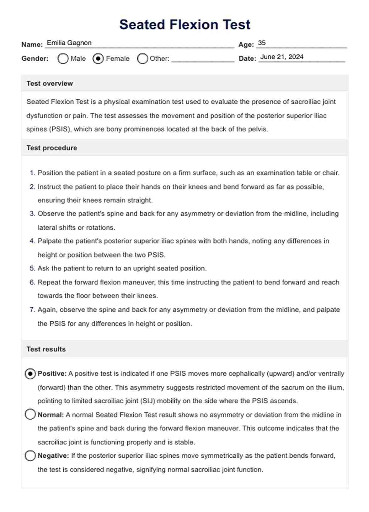
The Seated Flexion Test, also known as the sitting flexion test, is a physical examination technique used by healthcare practitioners to assess sacroiliac joint function and identify sacral dysfunctions.

During this test, the patient is seated with their feet flat on the floor and knees bent at a 90-degree angle. The examiner palpates the posterior superior iliac spines and asks the patient to bend forward slowly. The movement of the PSIS is observed; a positive test is indicated if one PSIS moves more superiorly than the other. This asymmetry suggests an issue with the sacral sulcus or sacral base.

The Seated Flexion Test is particularly useful for identifying extension dysfunctions and issues related to the oblique axis of the sacrum.

The test also helps in detecting inferior aspect or inferior portion dysfunctions by isolating the movement of the sacral base. During clinical tests, it is common for practitioners to perform both standing and seated flexion tests to compare results. Additionally, examining the patient prone can further validate findings and guide targeted interventions.

Next steps after conducting this test

After conducting the Seated Flexion Test and interpreting the results, healthcare practitioners typically proceed with several important next steps to further evaluate and manage the patient's condition. These steps are crucial for determining the appropriate treatment approach:

  1. Clinical correlation: The findings from the Seated Flexion Test are correlated with the patient's history, symptoms, and other physical examination findings. This helps to confirm the diagnosis and understand the extent of sacroiliac joint dysfunction.
  2. Additional tests and examinations: Depending on the initial findings, additional tests may be conducted to further assess sacroiliac joint stability, such as the standing flexion test, Gillet test, or Patrick's test. Imaging studies like X-rays or MRI scans may also be ordered to visualize the SI joints and rule out other possible causes of symptoms.
  3. Treatment planning: Based on the comprehensive assessment, a personalized treatment plan is developed. This may include osteopathic manipulative treatment (OMT), physical therapy, medication for pain management, or injections to alleviate inflammation in the SI joints.
  4. Patient education: Educating the patient about their condition, including lifestyle modifications and exercises to improve SI joint stability, is essential. Empowering patients with knowledge enhances their participation in their own care and promotes better outcomes.
  5. Follow-up: Regular follow-up appointments are scheduled to monitor progress, adjust treatment as needed, and ensure optimal recovery.

Benefits of conducting this test

Conducting the Seated Flexion Test offers several key benefits in clinical practice, aiding in the assessment and management of sacroiliac joint dysfunction:

Early detection of sacroiliac joint dysfunction

The Seated Flexion Test allows healthcare providers to detect subtle asymmetries in the movement of the posterior superior iliac spines early on. This early detection can prompt timely intervention and prevent the progression of sacroiliac joint dysfunction.

Differential diagnosis of low back pain

By assessing the sacral base and PSIS movements during the Seated Flexion Test, clinicians can differentiate between sacroiliac joint dysfunction and other causes of low back pain, such as lumbar spine issues or muscular imbalances. This aids in providing targeted treatment strategies based on accurate diagnosis.

Monitoring treatment progress

Repeat Seated Flexion Tests over time allow clinicians to monitor changes in PSIS movement and sacral alignment. Improved test results indicate progress in treatment, while persistent asymmetries may prompt adjustments in therapeutic approaches.

What is the Seated Flexion Test?

Commonly asked questions

What is the Seated Flexion Test?

The seated flexion test is a diagnostic technique used to assess sacroiliac joint dysfunction by observing asymmetry in the movement of the posterior superior iliac spines (PSIS) when a patient bends forward from a seated position.
What does a positive standing flexion test mean?

A positive standing flexion test indicates asymmetry or restriction in the motion of the sacroiliac joints when the patient bends forward from a standing position, suggesting dysfunction or imbalance in the pelvis or sacrum.

When performing a Seated Flexion Test the thumbs are placed?

When performing a Seated Flexion Test, the examiner places their thumbs on the posterior superior iliac spines (PSIS) to palpate and monitor the movement of these bony prominences during the patient's forward bending motion.

How do you assess for sacral torsion?

To assess for sacral torsion, the practitioner palpates for asymmetry or rotation in the sacrum compared to the ilia. This includes evaluating landmarks like the sacral base and PSIS to detect any rotational patterns indicative of sacral torsion.

Join 10,000+ teams using Carepatron to be more productive

One app for all your healthcare work