What is a PCL Anatomy Diagram?
A posterior cruciate ligament (PCL) anatomy diagram illustrates the anatomy of the PCL and provides a detailed visual representation essential for comprehending the ligament's role within the knee joint. On it, one can expect to see:
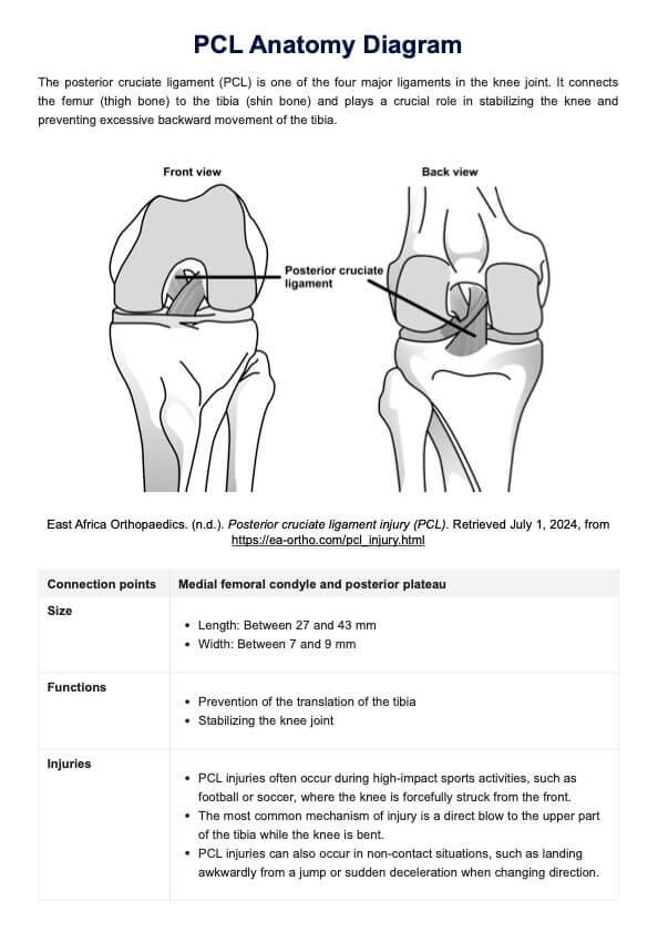
- Attachments of the PCL: The diagram illustrates the PCL's connection points, which are on the medial femoral condyle of the thigh bone and posterior plateau.
- PCL interactions: With the diagram, it's better clarified how the PCL interacts with other crucial instructions like the anterior cruciate ligament (ACL) and other ligaments in the joint structure, emphasizing its position and orientation.
- Shape and size of the PCL: The diagram outlines the shape and size of the PCL, underscoring its robust nature and the critical role it plays in preventing posterior tibial translation.
- Functions of the PCL: Annotations within the diagram often detail the PCL's functions, which include preventing the posterior translation of the tibia and being one of the two cruciate ligaments stabilizing the knee joint during knee flexion.
- PCL injuries: These visuals also address the significance of posterior cruciate ligament injuries, such as posterior cruciate ligament tears that can result from sports-related trauma or other knee ligament injuries.
- PCL treatment options: Surgical interventions like PCL reconstruction are frequently depicted to illustrate treatment options and rehabilitation strategies, reinforcing the diagram's utility in educational settings and clinical practice.
Do note that the content of a PCL Anatomy Diagram can differ depending on the creator, user, or source where one obtains a copy of the diagram


