


What is a stroke?
A stroke occurs when blood flow to a specific part of the brain is disrupted, either due to a blockage in a blood vessel (ischemic stroke) or when a blood vessel bursts (hemorrhagic stroke). This interruption deprives the brain tissue of oxygen and nutrients, leading to cell damage and potentially permanent neurological deficits. Among the expected consequences of acute stroke is the impairment of motor function, which affects muscle control and movement.
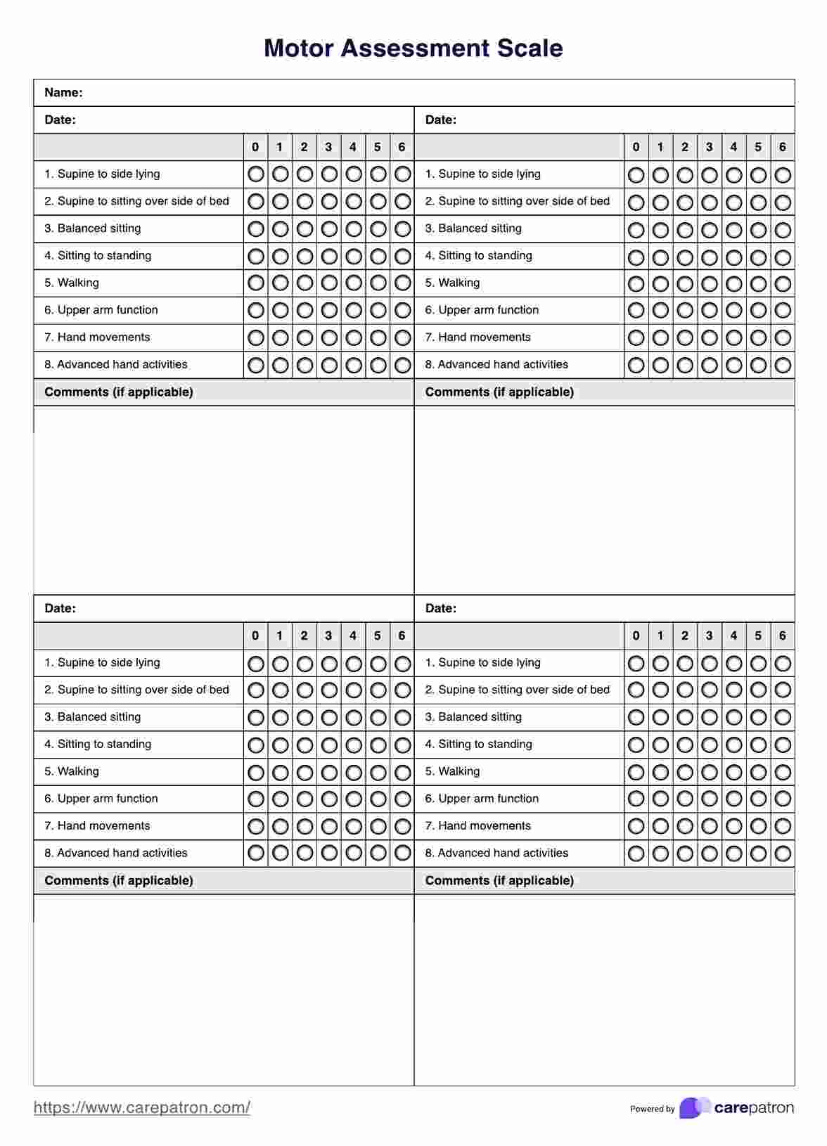
Assessing motor function post-stroke in clinical rehabilitation is paramount to designing effective treatment plans. Tools like the Action Research Arm Test (ARAT) are frequently employed to evaluate upper limb function, which is crucial for daily activities. Physical therapy, a cornerstone of stroke rehabilitation, focuses on restoring muscle strength, coordination, and range of motion.
Muscle tone, another aspect assessed during stroke rehabilitation, refers to the tension or resistance level in muscles at rest. Managing muscle tone aids in improving movement quality and function. Through physical medicine interventions and clinical rehab techniques, stroke patients undergo targeted therapies aimed at enhancing motor skills and overall functionality.
Reliability in stroke assessment tools ensures consistency in evaluating patient progress and treatment efficacy over time. With reliable measures, clinicians can track changes in motor function, muscle strength, and coordination, enabling tailored interventions to optimize recovery outcomes for stroke survivors.
How to diagnose a stroke
Diagnosing a stroke promptly is crucial for initiating appropriate treatment and minimizing long-term consequences. Here are the key steps and procedures involved:
- Assessment of symptoms: Recognize common signs of stroke such as sudden numbness or weakness in the face, arm, or leg, especially on one side of the body. Other symptoms include confusion, trouble speaking or understanding speech, difficulty seeing, dizziness, and severe headache.
- FAST test: Use the FAST acronym to assess for signs of stroke quickly: face dropping, arm weakness, speech difficulty, and time to call emergency services if all of these symptoms are present.
- Medical history review: Gather information about the patient's medical history, including risk factors such as hypertension, diabetes, smoking, and previous strokes or transient ischemic attacks (TIAs).
- Physical examination: Conduct a thorough neurological examination to assess motor function, sensation, coordination, and reflexes. Look for asymmetry or weakness in facial muscles, arm drift, and abnormal gait.
- Diagnostic tests: Perform imaging studies such as a CT scan or MRI to visualize the brain and identify areas of ischemia or bleeding. Additionally, a CT angiography or MR angiography may be used to evaluate blood flow in the cerebral arteries.
- Blood Tests: Analyze blood samples to assess for abnormalities such as elevated levels of glucose, lipids, or clotting factors, which may indicate an increased risk of stroke.
- Electrocardiogram (ECG or EKG): Record the electrical activity of the heart to detect arrhythmias or other cardiac abnormalities that could contribute to stroke risk.
- Carotid ultrasound: Use ultrasound imaging to assess for narrowing or blockages in the carotid arteries, a common cause of ischemic stroke.
- Lumbar puncture (spinal tap): Occasionally, a lumbar puncture may be performed to analyze cerebrospinal fluid for signs of bleeding or infection.


