Electrocardiogram

Understand your heart's health with an Electrocardiogram Test. This quick, painless procedure evaluates heart rhythm and detects potential heart conditions.

By Telita Montales on Jul 15, 2024.

tick

Fact Checked by Nate Lacson.

Use Template
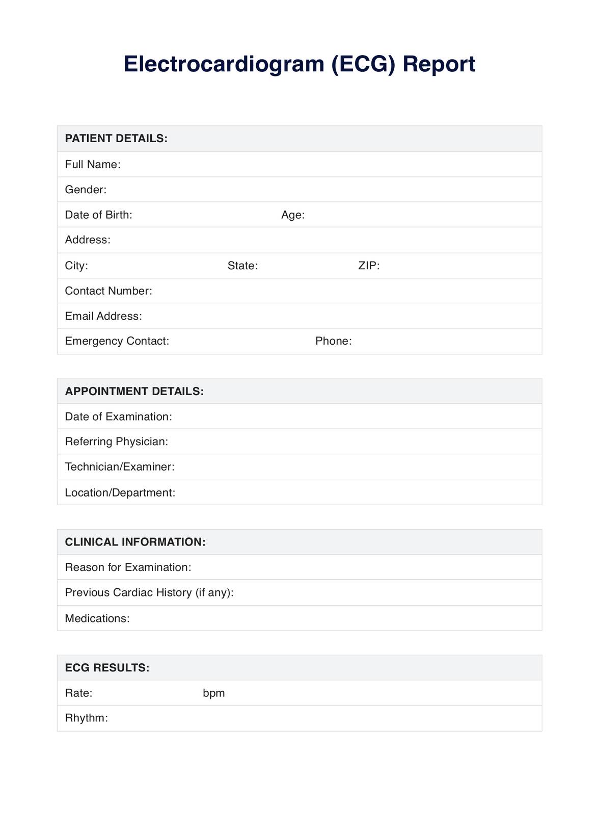
Electrocardiogram PDF Example
ToolbarShare uiAI Icon

What is an Electrocardiogram Test?

The electrocardiogram test, widely known as an EKG or ECG, is a key non-invasive diagnostic procedure that monitors the electrical activity pulsating through the heart. The importance of this test cannot be overstated, as it provides invaluable insights into the rhythm and rate at which the heart beats. It is a critical tool for healthcare providers in detecting and diagnosing various cardiac conditions, including arrhythmias (irregular heart rhythms), myocardial infarctions (heart attacks), and heart failure.

An EKG test involves attaching small electrode patches onto specific patient body areas. These areas typically include the chest, arms, and legs. The role of these electrodes is to serve as connectors, transmitting the electrical impulses generated by the heart to a recording machine.

This machine traces these signals, crafting a graph-like representation that depicts the heart's overall health and performance. This visual representation allows medical professionals to analyze the heart's condition and identify any potential abnormalities or issues that may be present.

We recommend watching this comprehensive explainer video to help you understand the intricate workings of an electrocardiogram test and its significance in diagnosing heart ailments. It provides a detailed walkthrough of the EKG test procedure and discusses its diagnostic importance.

For more extensive information about the EKG test, alongside other clinical tests, we invite you to visit our Electrocardiogram Tests section. Here, you will find a wealth of knowledge on various clinical tests, equipping you with the understanding you need to navigate your healthcare journey confidently.

How Does it Work?

Understanding how an electrocardiogram (EKG or ECG) test works is essential for healthcare professionals and patients. This knowledge helps ensure the test is conducted accurately and comfortably and results are interpreted correctly. Here's a step-by-step guide on how it works:

Step 1: Patient Preparation

The initial step in the EKG testing process involves preparing the patient for the procedure. This preparation stage includes explaining the entire process to the patient to alleviate fears or concerns about the test. The healthcare provider ensures the patient is comfortable and positions them correctly on the examination table, usually supine (lying face up). Comfort and understanding are key to ensuring the patient remains still during the test, which is crucial for obtaining accurate results.

Step 2: Placement of Electrodes

The next phase involves strategically placing approximately 10-12 electrode patches on the patient's body. These are typically placed on the skin of the chest, arms, and legs. The purpose of these electrodes is to detect the electrical activity generated by the heart, which forms the basis of the EKG test.

Step 3: Recording the ECG

Once the electrodes are correctly positioned and secured, they are connected to the ECG machine. This machine records the electrical impulses produced by the heart's rhythmic contractions and relaxations. These impulses are then visually displayed as wave patterns on a monitor, or they can be printed on paper for further examination.

Step 4: Interpretation of Results

The final step in the EKG process involves the interpretation of the recorded wave patterns. A trained healthcare professional examines these patterns to assess the heart's condition. They look for any wave abnormalities or irregularities that could indicate potential heart issues. The information gleaned from this interpretation is then used to guide the patient's further treatment and care.

We have prepared a printable Electrocardiogram Test for your convenience. This resource provides a visual guide to understanding the EKG process and interpreting the results.

When Would you use this Test?

The electrocardiogram (EKG or ECG) test is a crucial diagnostic tool in cardiology, but understanding when to use this test is essential for healthcare professionals and patients. It's not just a tool for cardiac specialists; it's also used by general practitioners, internists, and emergency medicine doctors.

An EKG test is primarily used when a healthcare provider suspects a patient may have heart-related problems. Symptoms often prompt an EKG test include chest pain, shortness of breath, dizziness, fainting, or rapid and irregular heartbeats. These symptoms might suggest conditions such as heart disease, arrhythmias, or heart attacks, which can be detected or confirmed by an EKG test.

However, an EKG test isn't only used when symptoms of heart problems are present. It's also a routine part of many health exams, especially for individuals with a family history of heart disease or other risk factors such as high blood pressure, diabetes, high cholesterol levels, or smoking. In these cases, an EKG test can help detect heart problems before symptoms appear, enabling early intervention and treatment.

Moreover, an EKG test is often conducted before surgeries, regardless of whether they're related to the heart. This pre-operative assessment helps determine the heart's condition and ability to withstand surgery's stress. It provides valuable information to anesthesiologists and surgeons, ensuring patient safety during and after the operation.

The electrocardiogram test is a versatile diagnostic tool that's used in various scenarios, from diagnosing heart conditions based on symptoms to routine health checks to pre-operative assessments. Its ability to provide real-time visual representations of the heart's electrical activity makes it invaluable in modern healthcare. Healthcare providers need to understand when using this resource is appropriate to ensure optimal patient care.

What do the Results Mean?

An electrocardiogram (EKG or ECG) test results are a wealth of information about the heart's condition. They offer insights into various aspects of heart health, including heart rate, rhythm, size, position, and more. However, understanding these results is crucial for appropriate diagnosis and treatment.

  • The heart rate is calculated based on the distance between each QRS complex, representing a single heartbeat. Adults' normal resting heart rate ranges from 60 to 100 beats per minute. Rates above or below this range could indicate tachycardia (rapid heart rate) or bradycardia (slow heart rate).
  • The heart's rhythm is determined by examining the consistency of the intervals between each QRS complex. An irregular rhythm, such as those seen in atrial fibrillation, can signify arrhythmia, a term used to describe abnormal heart rhythms.
  • The size and position of the heart can be inferred by looking at the magnitude and direction of the waveforms. For example, enlarged waves may suggest hypertrophy (enlargement) of the heart chambers, while changes in the typical waveform directions can indicate a shift in the heart's position within the chest.

Beyond these basic parameters, abnormalities found in the EKG waveforms can hint at various heart conditions. For instance, changes in the ST segment can indicate coronary artery disease or myocardial infarction (heart attack). Meanwhile, the presence of additional waves or missing waves can point towards conditions like bundle branch blocks or heart blocks.

EKG test results provide a comprehensive snapshot of the heart's electrical activity, offering valuable information about its health and function. However, interpreting these results requires expertise and should be done by a healthcare professional.

To help you understand these concepts better, we have a free Electrocardiogram Test for your reference. This resource provides a visual guide to understanding the different components of an EKG trace and their implications for heart health.

Research & Evidence

The electrocardiogram (ECG or EKG) has a rich history dating back to the early 20th century. The first practical ECG machine was developed in 1903 by Dutch physiologist Willem Einthoven, marking a significant milestone in cardiovascular medicine. Since then, the ECG has undergone numerous technological advancements, evolving into an essential diagnostic tool for heart diseases.

A vast body of research supports the effectiveness of the ECG in diagnosing various heart conditions. For instance, a study titled "The history, hotspots, and trends of electrocardiogram" discusses the broad applications of ECG in the clinical diagnosis and prognosis of cardiovascular disease (NCBI, 2015). Another study delves into the utility of ECG in differentiating normal from decreased systolic function in patients with heart failure (ScienceDirect, 2002).

Moreover, ECG interpretation accuracy is greatly influenced by the presentation of a patient's clinical history, as highlighted in a study published in Nature (2019). This underscores the importance of comprehensive patient history in conjunction with ECG for accurate diagnoses.

Furthermore, ECG is vital in predicting acute myocardial infarction, commonly known as a heart attack. A study on this topic emphasizes the importance of clinical history, examination, and ECG in the emergency room setting (ScienceDirect, 1991).

The electrocardiogram's historical development and extensive use in diagnosing and predicting heart conditions underscore its indispensable role in modern medicine. The wealth of research supporting its effectiveness testifies to its enduring relevance in healthcare.

References

  1. NCBI. (2015). The history, hotspots, and trends of electrocardiogram. Retrieved from https://www.ncbi.nlm.nih.gov/pmc/articles/PMC4554791/
  2. ScienceDirect. (2002). Utility of history, physical examination, electrocardiogram, and chest radiograph for differentiating normal from decreased systolic function in patients with heart failure. Retrieved from https://www.sciencedirect.com/science/article/pii/S0002934302010483
  3. Nature. (2019). Investigating the effect of clinical history before electrocardiogram interpretation on the visual behavior and interpretation accuracy of clinicians. Retrieved from https://www.nature.com/articles/s41598-019-47830-0
  4. ScienceDirect. (1991). Early prediction of acute myocardial infarction from clinical history, examination and electrocardiogram in the emergency room. Retrieved from https://www.sciencedirect.com/science/article/pii/0002914991907398
Who typically requests an Electrocardiogram Test?

Commonly asked questions

Who typically requests an Electrocardiogram Test?

Doctors typically request an ECG when they suspect heart disease or monitor patients' health with existing heart conditions.
When are Electrocardiogram Tests used?

ECGs are used in routine health exams, when symptoms suggest heart problems, or before surgeries to assess the heart's condition.

How are Electrocardiogram Tests used?

The test is used to measure the rate and regularity of heartbeats, the size and position of the heart chambers, and any damage to the heart.

How long does an Electrocardiogram Test take?

An ECG usually takes about 5 to 10 minutes.

Join 10,000+ teams using Carepatron to be more productive

One app for all your healthcare work