No items found.

Brain Imaging Techniques

Explore the latest advancements in brain imaging techniques. Learn about the various methods used to study the brain in detail.

By Wynona Jugueta on Feb 13, 2025.

Fact Checked by Gale Alagos.

Get Carepatron Free
Brain Imaging Techniques

What is brain imaging?

Brain imaging refers to using neuroimaging techniques to visualize and analyze the structure and function of the brain (Powers & Powers, 2014). These brain imaging methods are essential for diagnosing neurological disorders, assessing brain activity, and guiding treatment plans. They are widely used in cognitive neuroscience research and clinical settings to study brain areas, monitor disease progression, and evaluate therapeutic responses.

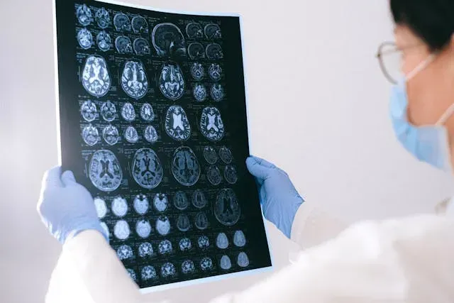
Brain imaging is divided into structural and functional Brain Imaging Techniques (Hirsch et al., 2015). Structural imaging, such as MRI and CT scans, provides detailed views of brain anatomy, helping detect conditions like tumors and stroke. Functional imaging, including fMRI and PET scans, assesses brain activity by measuring changes in brain waves, blood flow, or metabolic processes. These techniques are particularly useful for studying disorders like multiple sclerosis, where early detection of lesions can improve disease management.

Brain imaging advances our understanding of cognition, behavior, and neurological conditions by mapping the nervous system and tracking neural activity (Yen et al., 2023). These methods continue to evolve, improving diagnostic accuracy and enhancing treatment strategies for various brain disorders.

Purpose of brain imaging

Brain imaging's primary purpose is to diagnose and monitor brain-related conditions, such as tumors, strokes, and neurodegenerative diseases. It helps detect abnormalities in brain structure and function, assess the extent of damage following a brain injury, and evaluate the effectiveness of treatments.

Additionally, brain imaging is used in research to explore the neural basis of behavior, cognition, and emotion, contributing to the development of new therapies and interventions. This powerful tool aids clinicians in providing accurate diagnoses and personalized care for patients with various neurological and psychiatric disorders.

Brain Imaging Techniques

Brain Imaging Techniques are essential tools for visualizing the human brain (Xue et al., 2010). They help medical professionals diagnose neurological disorders, study specific brain regions, and guide treatments.

Structural imaging, such as CT and MRI, provides detailed anatomical views, while functional imaging, like fMRI and PET, captures brain activity in different brain regions. Advanced methods, including diffusion tensor imaging, enhance understanding of neural pathways. Below are key brain scanning techniques used in clinical and research settings.

Computed tomography (CT) scan

CT scans use X-rays to create cross-sectional images of the human brain (National Institute of Biomedical Imaging and Bioengineering, 2022). This technique is widely used for detecting tumors, strokes, and traumatic brain injuries. CT scans are quick and effective, making them ideal for emergency diagnostics. However, they expose patients to radiation, which limits repeated use.

Magnetic resonance imaging (MRI)

MRI provides high-resolution images of specific brain regions using strong magnetic fields and radio waves (National Institute of Biomedical Imaging and Bioengineering, n.d.). It is commonly used to diagnose tumors, multiple sclerosis, and brain atrophy. Unlike CT, MRI does not use radiation, making it safer for repeated scans.

Functional magnetic resonance imaging (fMRI)

fMRI measures changes in blood flow to analyze brain activity in different brain regions (Glover, 2011). This method is useful for studying cognitive functions and identifying abnormalities related to psychiatric and neurological disorders. It is widely used in both clinical and research settings.

Positron emission tomography (PET)

PET scans use radioactive tracers to detect metabolic activity in the brain (Kapoor & Kasi, 2022). This technique helps assess conditions like Alzheimer’s disease, epilepsy, and cancer by identifying changes in glucose metabolism. PET is often combined with CT or MRI for a more comprehensive evaluation.

Electroencephalography (EEG)

EEG records electrical activity in the brain by detecting brain wave patterns (Abhang et al., 2016). It is commonly used to diagnose epilepsy, sleep disorders, and brain function abnormalities. EEG is a non-invasive and cost-effective tool for monitoring real-time brain activity.

Magnetoencephalography (MEG)

MEG measures magnetic fields produced by neuronal activity, offering high temporal resolution (Singh, 2014). It is primarily used for mapping brain functions before surgery and diagnosing epilepsy. MEG provides more precise data than EEG but is less widely available due to its high cost.

Single-photon emission computed tomography (SPECT)

SPECT imaging evaluates cerebral blood flow using radioactive tracers (Yandrapalli & Puckett, 2022). It helps detect conditions like stroke, dementia, and brain injuries. While similar to PET, SPECT is more cost-effective but provides lower-resolution images.

Event-related optical signal (EROS)

EROS is an emerging technique using infrared light to measure brain activity in specific regions (Gratton & Fabiani, 2001). It offers high temporal resolution, making it useful for studying cognitive functions without radiation exposure.

Functional ultrasound imaging (fUSI)

fUSI is a relatively new technique that uses ultrasound waves to monitor neuroimaging techniques related to brain activity (Martinez de Paz & Macé, 2021). It provides high spatial and temporal resolution, making it useful for brain function and neural connectivity research.

What should you consider when choosing a brain imaging technique?

Selecting the right brain imaging technique depends on the clinical need, patient condition, and specific diagnostic requirements. An MRI machine provides detailed anatomical images, while PET and fMRI are better for assessing brain function. Each technique offers unique insights, making the choice dependent on the investigated condition.

Practical factors like patient safety, cost, and accessibility also play a role. MRI may not be suitable for those with metal implants, and PET scans can be costly and less available compared to CT scans. Balancing these considerations ensures the most effective imaging method is used.

Who can benefit from knowing the Brain Imaging Techniques?

Brain Imaging Techniques are essential for healthcare professionals and researchers. Neurologists rely on CT and MRI scans to diagnose conditions like stroke, epilepsy, and dementia by identifying structural abnormalities. Radiologists interpret brain images to detect tumors, infections, and injuries, aiding in precise diagnoses.

Psychiatrists use functional MRI (fMRI) to study brain activity changes in mental health disorders like depression and schizophrenia, improving treatment strategies.

Researchers also benefit from these techniques to analyze brain structure and function in healthy individuals and those with neurological conditions. Their findings contribute to a deeper understanding of the brain and the development of new treatments.

References

Abhang, P. A., Gawali, B. W., & Mehrotra, S. C. (2016). Technological basics of EEG recording and operation of apparatus. In Introduction to EEG- and speech-based emotion recognition (pp. 19–50). Academic Press. https://doi.org/10.1016/b978-0-12-804490-2.00002-6

Gratton, G., & Fabiani, M. (2001). The event-related optical signal: A new tool for studying brain function. International Journal of Psychophysiology, 42(2), 109–121. https://doi.org/10.1016/s0167-8760(01)00161-1

Glover, G. H. (2011). Overview of functional magnetic resonance imaging. Neurosurgery Clinics of North America, 22(2), 133–139. https://doi.org/10.1016/j.nec.2010.11.001

Hirsch, G. V., Bauer, C. M., & Merabet, L. B. (2015). Using structural and functional brain imaging to uncover how the brain adapts to blindness. Annals of Neuroscience and Psychology, 2, 5. https://pmc.ncbi.nlm.nih.gov/articles/PMC6168211/

Kapoor, M., & Kasi, A. (2022, October 3). PET scanning. StatPearls Publishing. https://www.ncbi.nlm.nih.gov/books/NBK559089/

Martinez de Paz, J. M., & Macé, E. (2021). Functional ultrasound imaging: A useful tool for functional connectomics? NeuroImage, 118722. https://doi.org/10.1016/j.neuroimage.2021.118722

National Institute of Biomedical Imaging and Bioengineering. (n.d.). Magnetic resonance imaging (MRI). National Institute of Biomedical Imaging and Bioengineering. https://www.nibib.nih.gov/science-education/science-topics/magnetic-resonance-imaging-mri

National Institute of Biomedical Imaging and Bioengineering. (2022, June). Computed tomography (CT). National Institute of Biomedical Imaging and Bioengineering. https://www.nibib.nih.gov/science-education/science-topics/computed-tomography-ct

Powers, K. E., & Powers, W. J. (2014). Brain imaging, physiological. In Encyclopedia of the neurological sciences (pp. 486–488). Academic Press. https://doi.org/10.1016/B978-0-12-385157-4.00202-5

Singh, S. P. (2014). Magnetoencephalography: Basic principles. Annals of Indian Academy of Neurology, 17(5), 107. https://doi.org/10.4103/0972-2327.128676

Xue, G., Chen, C., Lu, Z.-L., & Dong, Q. (2010). Brain imaging techniques and their applications in decision-making research. Acta Psychologica Sinica, 42(1), 120–137. https://doi.org/10.3724/sp.j.1041.2010.00120

Yen, C., Lin, C.-L., & Chiang, M.-C. (2023). Exploring the frontiers of neuroimaging: A review of recent advances in understanding brain functioning and disorders. Life, 13(7), Article 1472. https://doi.org/10.3390/life13071472

Commonly asked questions

Is MRI or EEG better for the brain?

MRI is better for detailed brain structural imaging, while EEG excels at detecting real-time electrical activity and brain wave patterns. The choice depends on the clinical need—MRI for anatomical evaluation and EEG for functional assessments like epilepsy monitoring.

What are the different techniques of brain imaging?

Brain imaging includes structural methods like CT and MRI and functional methods such as fMRI, PET, EEG, MEG, SPECT, EROS, and fUSI. These neuroimaging techniques help visualize specific brain regions, assess different brain regions, and track brain function or abnormalities.

What is the most effective brain imaging technique?

The most effective technique depends on the purpose—MRI provides high-resolution structural images, while fMRI, PET, and EEG are best for functional assessments. For mapping neural pathways, diffusion tensor imaging (DTI) is highly effective in studying white matter integrity.

Join 10,000+ teams using Carepatron to be more productive

One app for all your healthcare work