What are the Ottawa Knee Rules?
The Ottawa Knee Rules are clinical decisions designed to help healthcare professionals decide when knee radiographs are needed in acute knee injury cases. These guidelines minimize unnecessary radiography by identifying specific criteria that indicate high-risk injuries and clinically significant fractures. By accurately identifying patients who need imaging, the Ottawa Knee Rules reduce radiation exposure and optimize resources and use of radiography in emergency departments, improving overall clinical practice and patient care.
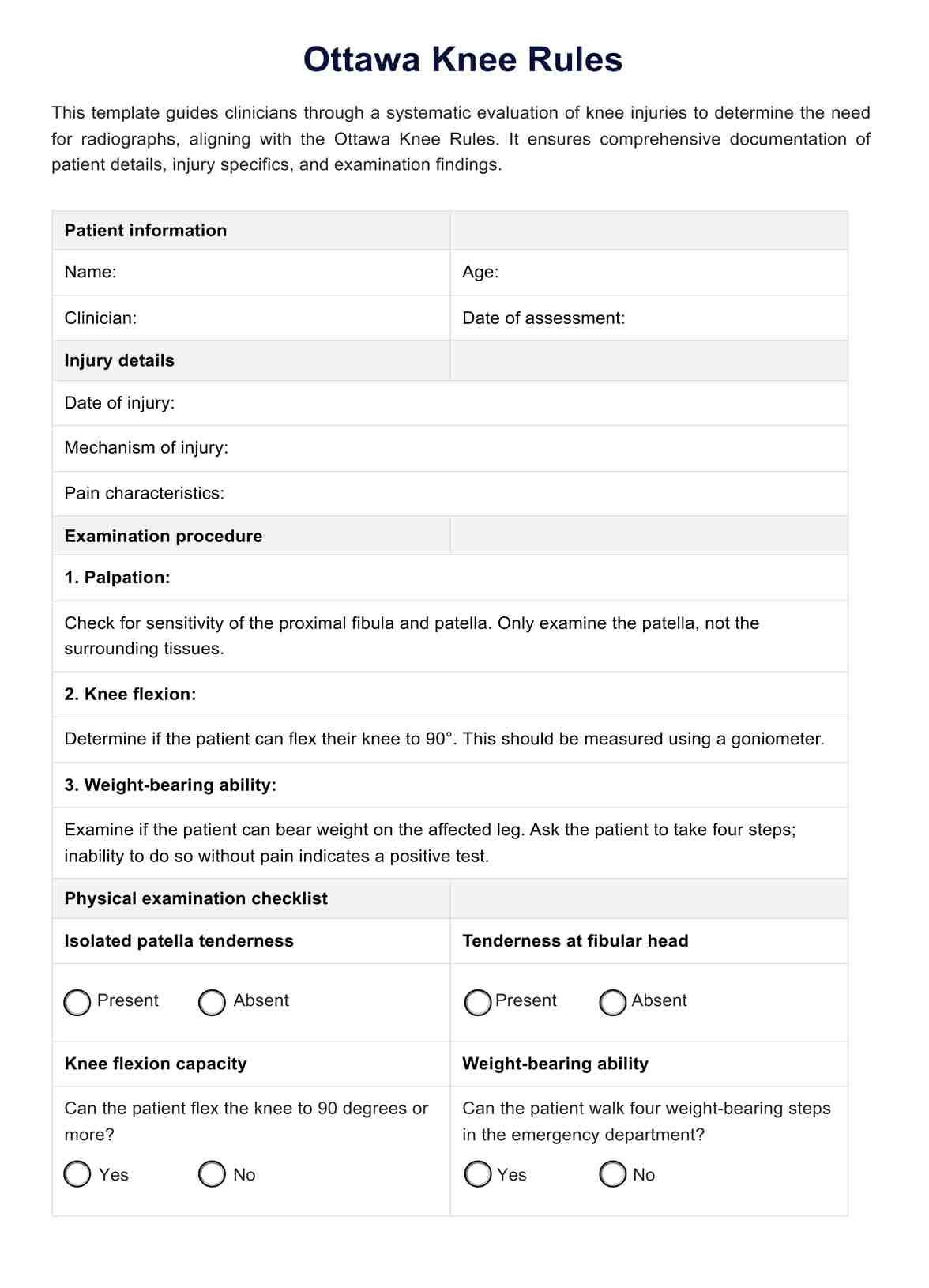
Ottawa Knee Rules criteria
The Ottawa Knee Rules suggest that radiography in acute knee is only needed if any of the following criteria are met:
- Age 55 or older
- Tenderness at the head of the fibula
- Isolated tenderness of the patella
- Inability to flex the knee to 90 degrees
- Inability to bear weight both immediately and in the emergency department (four steps; unable to transfer weight twice onto each leg regardless of limping)
How are the Ottawa Knee Rules applied?
The Ottawa Knee Rules are applied through a systematic examination procedure that helps clinicians accurately determine whether a patient with acute knee trauma requires radiographic imaging. This approach ensures efficient use of resources in emergency departments and reduces unnecessary radiation exposure. Here’s a step-by-step guide on how the rules are applied:
Patient interview
Begin by conducting a quick interview with the patient to understand the mechanism of injury and how it occurred. For instance, was the knee injury caused by blunt trauma from a fall, sports incident, or direct blow? Inquire about any history of prior knee injuries or surgeries that could affect the current assessment. Have the patient describe the intensity and nature of their knee pain, noting whether it worsens with specific movements or positions.
Physical examination
A comprehensive physical examination of the knee begins with palpating to locate specific areas of pain or tenderness. This should include checking for isolated tenderness directly over the patella or kneecap and at the fibula's head, just below the knee joint on the lateral side. Both areas are critical for identifying potential injuries or disorders.
Next, assessing the range of motion is crucial for determining the knee's functional capacity. Unlike tenderness, range of motion should be measured using a goniometer to obtain an accurate reading. Position the goniometer at the lateral aspect of the knee, aligning it with the femur and the fibula. Ask the patient to flex and extend the knee. A normal knee should flex to at least 90 degrees without significant pain. Difficulty achieving this range or experiencing pain during the movement is a critical indicator that may require further imaging to identify potential internal damage or structural issues.
Assessment of walking ability
Evaluate the patient’s ability to bear weight and walk unassisted immediately after the injury and within the emergency department. The patient should be able to transfer weight at least twice onto each leg and take four weight-bearing steps without excessive limping or collapsing.
By following this detailed examination procedure using the Ottawa Knee Rules, clinicians can effectively identify high-risk knee injuries that warrant radiographs. This standardized approach helps to differentiate between clinically significant fractures and less severe injuries that can be managed conservatively.