


What is a distal radius fracture?
A distal radius fracture is a break near the wrist end of the radius, one of the two main bones in the forearm. It is one of the most common types of fractures, particularly affecting older adults due to osteoporosis, as well as in early age and children through high-impact activities or trauma, such as falls or sports injuries. These fractures can significantly impact wrist function and require precise diagnosis and treatment to ensure proper healing.
Symptoms
Distal radius fractures present with various symptoms that can impact daily activities and overall wrist function.
- Immediate pain at the fracture site, often accompanied by swelling and bruising.
- Visible deformity or an abnormal bend in the wrist.
- Difficulty or inability to move the wrist or fingers.
- Tenderness to touch near the site of the break.
Types of distal fractures
Understanding the different types of distal radius fractures helps in accurate diagnosis and appropriate treatment planning.
- Colles' fracture: A fracture of the distal radius with dorsal displacement of the wrist and hand, typically caused by falling onto an outstretched hand.
- Smith's fracture: Also known as a reverse Colles' fracture, it involves a volar displacement of the distal fragment, usually resulting from a fall onto a flexed wrist.
- Barton's fracture: An intra-articular fracture of the distal radius with dislocation of the radiocarpal joint, often due to a direct blow to the wrist or a high-energy impact.
- Hutchinson's fracture: Also known as a chauffeur's fracture, this is a fracture of the radial styloid process, commonly caused by a direct blow to the wrist or a twisting injury.
Other types include:
- Intra-articular fractures: These extend into the wrist joint, complicating treatment and recovery.
- Extra-articular fractures: Fractures that do not involve the joint surface.
- Comminuted fractures: The bone is broken into multiple pieces.
- Open fractures: The bone breaks through the skin, requiring urgent medical attention due to infection risk
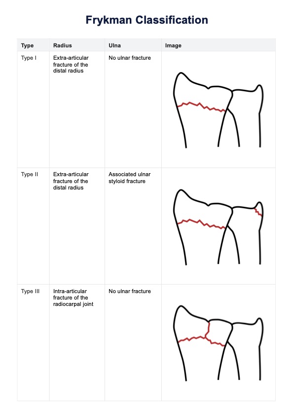
How to classify fractures
Classifying fractures involves assessing the specific location and nature of the break. Various classification systems exist to categorize fractures based on these criteria. Healthcare professionals use radiographic imaging to determine the exact type and severity of the fracture, aiding in the selection of the appropriate treatment approach. Systems like the Frykman Classification are instrumental in providing a structured framework for these assessments.


