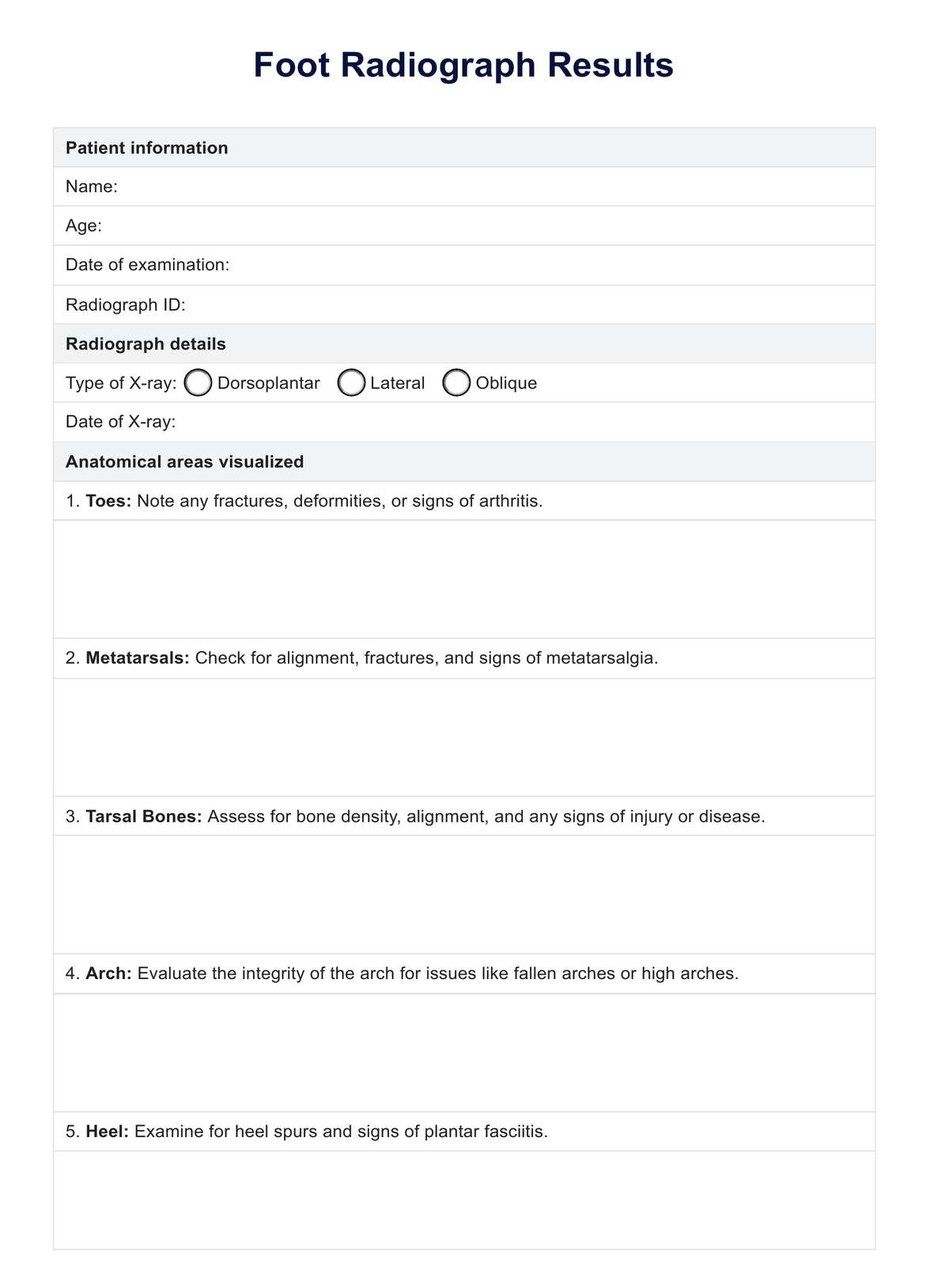
What can Foot Radiographs be used for?
Foot radiographs, commonly known as foot X-rays, are a fundamental tool in medical imaging that serve a variety of set of diagnostic and assessment purposes in healthcare:
Detection of fractures and bone injuries
X-rays are primarily used to visualize breaks in the bones of the foot, such as fractures from falls, sports injuries, or accidents. They can also identify smaller bone injuries like chips or hairline fractures that might not be immediately apparent.
Assessment of bone alignment and deformities
Radiographs provide clear images of the positioning and alignment of the bones within the foot. This is essential for diagnosing conditions like flat feet, high arches, or more complex deformities affecting walking and balance.
Diagnosis of common foot pathologies
X-rays can diagnose various foot conditions, including bunions, arthritis, and heel spurs. These conditions often affect mobility and comfort, making X-rays a valuable tool for planning treatment.
Evaluation of soft tissue conditions
While X-rays are primarily used to view bones, they can also assess soft tissue conditions, especially those involving calcifications or chronic diseases.
Monitoring disease progression and treatment efficacy
For chronic conditions or after surgical interventions, foot radiographs help monitor changes over time, assess the effectiveness of treatments, and plan future interventions.
Pre-surgical planning
Before foot surgery, detailed X-rays can help surgeons plan the procedure by providing a clear map of bone and joint structures.
Radiographs offer a detailed view of the foot's internal structure and play a crucial role in medical diagnostics. They guide the treatment and management of various conditions affecting the foot, toes, and lower extremities.