Posterior Sag Sign
Detect posterior cruciate ligament injuries accurately with the Posterior Sag Sign, a reliable physical examination maneuver.

What is a posterior cruciate ligament tear?
A posterior cruciate ligament (PCL) tear is a significant knee injury, often resulting from sudden trauma or force applied to the knee. The PCL is located behind the anterior cruciate ligament (ACL) and helps stabilize the knee by preventing excessive backward movement of the tibia relative to the femur. Alongside other structures like the fibular collateral ligament, the PCL maintains proper alignment and function of the knee joint.
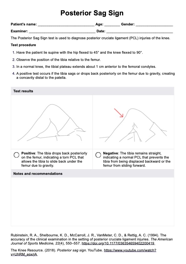
One key physical examination finding for diagnosing a PCL tear is the Posterior Sag Sign, in which the tibia sags backward when the knee is flexed. This sign is both specific and sensitive for detecting posterior cruciate ligament injuries. Tests like the posterior drawer and Lachman tests may also be used to confirm the diagnosis.
In more severe cases, a torn PCL can lead to additional complications, especially during surgical reconstruction. For instance, there is a risk of popliteal artery injury, which can significantly affect recovery and overall knee function. These potential complications underscore the importance of accurate diagnosis and thorough treatment planning for PCL tears.
Symptoms of this tear
A posterior cruciate ligament tear manifests through several key symptoms that can significantly affect daily activities and overall knee stability. In cases of isolated PCL injuries, symptoms may appear more subtle if other knee structures remain intact. Nonetheless, these injuries can still lead to discomfort and hinder normal function.
- Pain
- Instability
- Swelling
- Limited mobility
- Tenderness
What can tear posterior cruciate ligaments?
PCL tears can occur due to several common causes. Understanding these can help with prevention and treatment. The main causes include:
- Direct impact
- Sports injuries
- Hyperextension
- Hyperflexion
- Rotational forces
- Combined injuries
Posterior Sag Sign Template
Posterior Sag Sign Example
What is the Posterior Sag Sign?
The Posterior Sag Sign is a clinical test for diagnosing a posterior cruciate ligament injury. As mentioned, it involves observing the position of the tibia (shinbone) in relation to the femur (thighbone) when the knee is bent. The assessment of the medial femoral condyle in the Posterior Sag Sign serves as a visual indicator of PCL laxity or injury. A noticeable posterior sag of the tibia relative to the femur, accompanied by a depression of the medial femoral condyle, suggests a positive test result and indicates potential PCL pathology. This observation guides further diagnostic evaluation and treatment planning for individuals suspected of having PCL injuries.
How is this assessment conducted?
The patient is lying on their back with their hips flexed and knees bent to 90 degrees, known as the "tabletop" position. The examiner observes the patient's knee profile from the side and looks for the posterior displacement of the tibia in relation to the femur. A positive Posterior Sag Sign indicates that the PCL is not functioning properly, allowing the tibia to slide backward.
To confirm a PCL tear, the examiner may perform tests like the posterior drawer test, where the patient's knee is bent to 90 degrees and the tibia is pushed backward. A positive test occurs if the tibia moves excessively backward, indicating a PCL tear.
For easy and consistent assessment, click the "Use template" button on this page. Our template contains the procedure and some images to guide you in determining whether the Posterior Sag Sign test is positive or not.
Benefits of using our test template
Using our Posterior Sag Sign template offers several benefits in the evaluation of PCL injuries, facilitating precise diagnosis and targeted management strategies to optimize patient outcomes. Check these out:
- Streamlined documentation: The template provides clear fields for recording each test detail, making it easier to note findings and track outcomes over time.
- Accurate assessment of acute PCL injuries: By organizing key observations, the template helps healthcare professionals quickly identify signs of acute PCL injuries, facilitating timely diagnosis and intervention.
- Improved communication: Standardized fields ensure clear, consistent documentation, enhancing collaboration among multiple clinicians involved in the patient’s care.
- Efficient follow-up: The template’s structured layout makes it easy to compare current findings with previous results, allowing practitioners to evaluate progress or detect new concerns promptly.
- Enhanced patient education: Providing a tangible record of test results helps patients understand their condition, reinforcing adherence to treatment plans and follow-up appointments.
Commonly asked questions
A posterior sag refers to the visible drop of the tibia behind the femur when the knee is flexed at 90 degrees. This often signals a potential injury to the posterior cruciate ligament, which normally prevents excessive backward movement of the tibia.
A positive Posterior Sag Sign suggests the proximal tibia shifts backward during knee flexion, indicating possible posterior cruciate ligament insufficiency. Posterior tibial displacement points to compromised PCL stability, allowing the tibia to sag relative to the femur.
In some mild cases, especially with partial tears, conservative management (like bracing and physical therapy) may help the PCL heal. However, complete tears often require surgical intervention to restore full knee stability and function.
Yes, other tests than the posterior sag test include the posterior drawer test, MRI imaging, and arthroscopy. These tests help confirm the diagnosis and assess the severity of the injury.