Myasthenia Gravis (MG) Test

Accurate Myasthenia Gravis (MG) Test for precise diagnosis and treatment planning. Trust our healthcare expertise for reliable results and personalized care.

By Bernard Ramirez on May 09, 2024.

Fact Checked by RJ Gumban.

Use Template

What are myasthenia gravis (MG) tests?

Myasthenia Gravis (MG) tests are pivotal tools for healthcare practitioners in diagnosing and managing this neuromuscular disorder. These tests provide precise insights into a patient's condition and guide practitioners toward tailored treatment strategies.

The primary diagnostic tests for MG include the Edrophonium test, blood tests for acetylcholine receptor antibodies, and repetitive nerve stimulation. These tests collectively aid in confirming the presence of MG, assessing its severity, and informing treatment decisions.

The Edrophonium test involves administering a short-acting medication to temporarily alleviate muscle weakness, helping practitioners observe a positive response in MG patients. Blood tests check for specific antibodies, aiding in definitive diagnosis.

Repetitive nerve stimulation is another essential test, evaluating how well nerves can repeatedly send signals to muscles. In MG, impaired signal transmission leads to muscle weakness and fatigue, a key aspect assessed through this test.

These tests empower healthcare practitioners to design personalized care plans, ensuring optimal outcomes for MG patients. With accurate diagnostics, practitioners can tailor treatments, combining medication, lifestyle adjustments, and, in some cases, surgical interventions.

Navigating MG management becomes a collaborative journey between practitioners and patients, fostering a positive and supportive healthcare environment. As we delve into Myasthenia Gravis testing, let's embrace the power of knowledge to enhance patient care and inspire hope in this challenging condition.

The journey towards a conclusive MG diagnosis initiates with a meticulous clinical evaluation. Practitioners delve into the patient's medical history, exploring symptoms such as muscle weakness, fatigue, and ptosis, a critical step in identifying potential indicators of MG.

A cornerstone in MG diagnosis is blood testing, specifically targeting acetylcholine receptor (AChR) and muscle-specific tyrosine kinase (MuSK) antibodies. These antibodies are a critical marker, indicating an autoimmune basis for the neuromuscular dysfunction observed in MG patients.

Neurophysiological testing, including nerve conduction studies and electromyography, delves into the electrical activity of muscles and nerves. This aids in pinpointing neuromuscular transmission defects characteristic of MG, contributing to a comprehensive understanding of the disease.

Imaging techniques such as CT scans and MRI play a vital role, particularly in visualizing affected muscles. This provides a tangible representation of the extent and localization of muscle abnormalities, aiding practitioners in refining their diagnosis and treatment plans.

The ice test is a unique diagnostic tool involving the application of a cold stimulus to specific muscle groups. In MG patients, this temporary improvement in muscle strength helps confirm the neuromuscular dysfunction, contributing to the overall diagnostic puzzle.

MG often manifests in facial muscles and eye movements, so a thorough clinical examination of these areas is paramount. Observing the response of the eye muscles to various stimuli and assessing facial muscle strength aids in differentiating between ocular and generalized MG.

During various tests, including nerve conduction studies and imaging, monitoring vital signs such as blood pressure and respiratory function ensures the safety and well-being of MG patients throughout the diagnostic process.

Enzyme-linked immunosorbent Assay (ELISA) is a laboratory test used to detect antibodies, including AChR antibodies, further solidifying the autoimmune etiology of MG. This precise methodology enhances the specificity of the diagnosis.

Myasthenia Gravis tests encompass a multidimensional approach, combining clinical evaluation, blood tests, neurophysiological studies, imaging, and specialized diagnostic tools. This comprehensive strategy enables healthcare practitioners to unravel the intricacies of MG, facilitating accurate diagnoses and personalized treatment plans for adult patients grappling with this autoimmune neuromuscular disorder.

Printable Myasthenia Gravis (MG) Test

Download this Myasthenia Gravis (MG) Test for healthcare practitioners in diagnosing and managing this neuromuscular disorder.

Testing

Myasthenia Gravis (MG) is a complex neuromuscular disorder requiring meticulous testing for accurate diagnosis and effective management. The journey begins with a comprehensive medical history and physical examination. Practitioners keenly observe symptoms such as muscle weakness, fatigue, and ptosis, especially in ocular MG, where the eye muscles are predominantly affected.

Laboratory tests, including blood tests that assess the presence of acetylcholine receptor (AChR) antibodies, are crucial to the diagnostic process. These antibodies are pivotal in disrupting neuromuscular transmission, leading to muscle weakness in MG patients. The detection of AChR antibodies through enzyme-linked immunosorbent assay (ELISA) provides valuable insights into the autoimmune nature of MG.

Blood tests

One of the primary tools in the diagnostic arsenal is blood testing. This involves screening for acetylcholine receptor (AChR) antibodies, a hallmark in most MG patients. These antibodies indicate an autoimmune response targeting the acetylcholine receptors, disrupting neuromuscular transmission and leading to muscle weakness.

Edrophonium (ice pack) test

The Edrophonium or Ice Pack Test is another dynamic diagnostic method. It involves administering a short-acting medication or applying an ice pack to alleviate muscle weakness in MG patients temporarily. A positive test result signifies a transient improvement in muscle strength, confirming MG's neuromuscular transmission defect characteristic.

Imaging studies

Magnetic Resonance Imaging (MRI) and Computed Tomography (CT) scans are employed to visualize and assess the affected muscles and nerves. These imaging studies contribute valuable insights into disease severity and aid in ruling out other neuromuscular disorders.

Nerve conduction study

Practitioners may conduct a nerve conduction study to evaluate nerve and muscle function. This test measures the electrical activity in muscles and nerves, helping identify the neuromuscular transmission abnormalities indicative of MG.

Clinical examination

A thorough physical examination is crucial in the diagnosis of MG. Practitioners assess muscle tone, strength, and weakness, particularly focusing on muscles commonly affected, such as those controlling eye movements, facial expressions, and swallowing.

Medical history:

Understanding the patient's medical history is integral. Detailed information on symptoms, their onset, and any family history of autoimmune disorders provides essential context for a comprehensive diagnosis.

Seronegative MG and musk antibodies

Sometimes, patients may present with seronegative MG, lacking detectable AChR antibodies. Additional testing for muscle-specific tyrosine kinase (MuSK) antibodies becomes imperative in such scenarios, aiding in diagnosing this specific subtype.

Monitoring treatment response

Beyond initial diagnosis, these tests are vital in monitoring treatment response. Adjustments in medication and other interventions can be tailored based on the evolving needs of MG patients.

Navigating the intricacies of MG testing requires a multidimensional approach, leveraging the synergy of blood tests, imaging studies, and clinical assessments. As healthcare practitioners engage in this diagnostic journey, the focus remains on delivering precise and personalized care, instilling hope and empowerment in MG patients as they embark on the path to improved health and well-being.

Laboratory tests

Laboratory tests stand at the forefront of Myasthenia Gravis diagnostics. Blood tests are pivotal in detecting specific antibodies like AChR antibodies and muscle-specific tyrosine kinase (MuSK) antibodies. Positive results in these tests provide a concrete foundation for diagnosing autoimmune MG, guiding practitioners toward tailored treatment plans.

Sometimes, patients may present with seronegative MG, where traditional blood tests yield negative results. Here, additional tests, such as the ice pack test, come into play. This unique diagnostic tool involves applying a cold pack to affected muscles, temporarily improving muscle strength in MG patients, thereby indicating a positive test result.

Electrodiagnostic testing

Electrodiagnostic testing is pivotal in comprehensively evaluating patients with myasthenia gravis (MG). Practitioners gain valuable insights into the neuromuscular junction and muscle function by utilizing various tests such as repetitive nerve stimulation, nerve conduction studies, and single-fiber electromyography.

Repetitive nerve stimulation involves the delivery of rapid nerve impulses, assessing the decremental response in muscle contraction characteristic of MG. Nerve conduction studies evaluate nerve impulses' speed and efficiency, aiding in identifying abnormalities in neuromuscular transmission. Single-fiber electromyography provides detailed insights into the function of individual muscle fibers, pinpointing specific neuromuscular transmission defects.

Pharmacological testing

Pharmacological testing is a crucial component in the diagnostic journey of Myasthenia Gravis (MG) patients, confirming the presence of this neuromuscular disorder and guiding treatment decisions. The Edrophonium test, a hallmark of pharmacological testing, involves temporarily administering a short-acting medication to improve muscle strength. This test is precious in cases where the clinical presentation is ambiguous, providing a rapid and observable response.

The ice pack test is another pharmacological approach to evaluating the impact of cooling on muscle weakness. By applying a cold pack to specific muscle groups, practitioners can assess improvements or worsening of symptoms, contributing to the diagnostic process.

What happens during MG testing?

Myasthenia Gravis (MG) testing is a systematic process designed to uncover the intricate details of this neuromuscular disorder. The journey begins with a thorough medical history and a neurological examination, where practitioners assess muscle weakness, double vision, and other symptoms that may indicate MG.

Blood tests play a pivotal role in MG diagnosis. These include AChR antibody testing, revealing the presence of antibodies attacking acetylcholine receptors, and MuSK antibody testing for specific subsets of MG patients. Striated muscle antibodies contribute to diagnostic sensitivity, aiding in identifying seronegative MG cases.

Electrodiagnostic testing follows, incorporating repetitive nerve stimulation and nerve conduction studies. These procedures delve into the neuromuscular junction, evaluating abnormal neuromuscular transmission and muscle contraction. Single-fiber electromyography further refines the assessment, pinpointing individual muscle fibers affected by MG.

Pharmacological testing involves iconic procedures such as the Edrophonium and ice pack tests. The Edrophonium test, utilizing a short-acting medication, assesses rapid improvements in muscle strength, aiding in confirming the diagnosis. The ice pack test, applied to specific muscle groups, evaluates the impact of cooling on muscle weakness, providing additional diagnostic insights.

Myasthenia Gravis test example (sample)

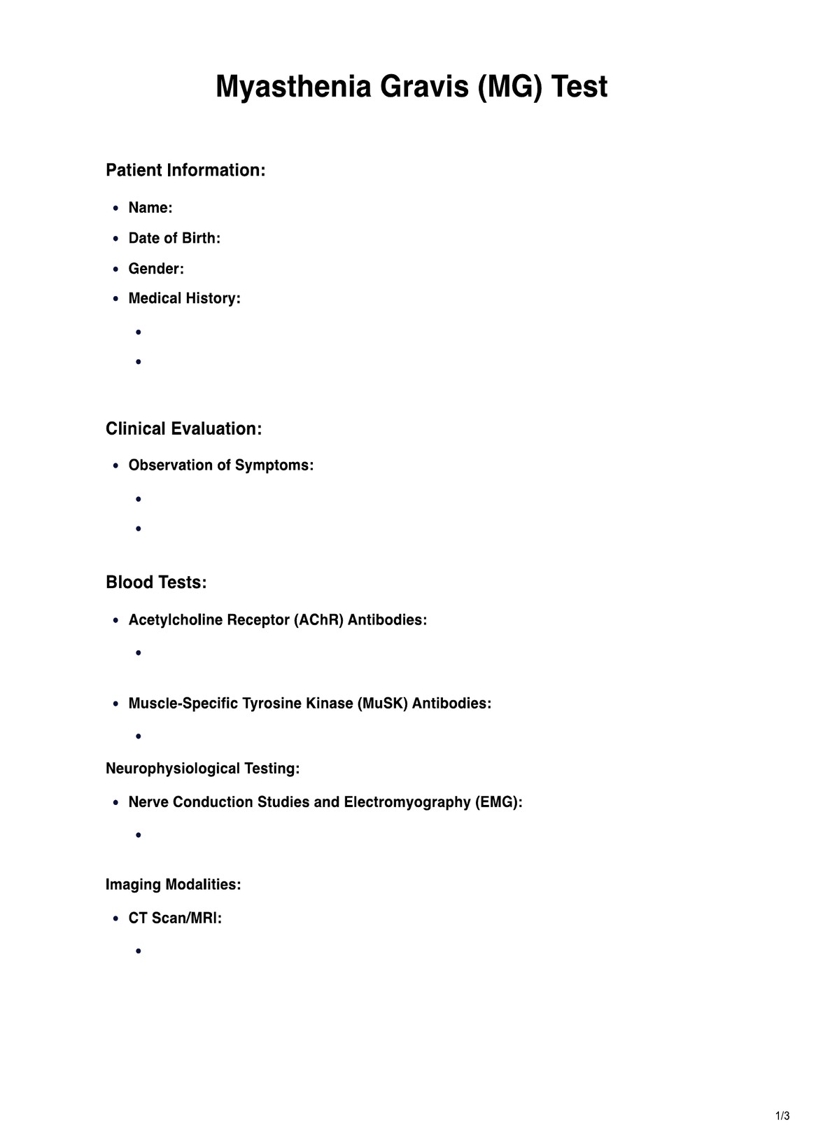
The Myasthenia Gravis (MG) Testing Template is a comprehensive document for healthcare practitioners managing MG patients. It integrates essential elements such as medical history, presenting symptoms, and detailed results from AChR antibody tests, electrodiagnostic procedures, and pharmacological tests like the Edrophonium and ice pack tests. Imaging studies, including CT scans, are incorporated to assess thymus gland involvement.

The template emphasizes a collaborative approach, guiding practitioners to diagnose MG, monitor treatment responses, and make informed decisions for personalized patient care. This dynamic tool ensures meticulous documentation and facilitates ongoing assessments in managing this complex neuromuscular disorder.

Download Myasthenia Gravis (MG) Test example:

Myasthenia Gravis (MG) Test example

Antibodies in diagnosis of Myasthenia Gravis

Antibody testing is pivotal in diagnosing Myasthenia Gravis (MG), particularly in assessing acetylcholine receptor (AChR) antibodies. Blood tests specifically targeting AChR antibodies provide crucial insights into the autoimmune nature of MG. Elevated levels of AChR antibodies indicate an immune system attack on acetylcholine receptors, impairing neuromuscular transmission. In conjunction with clinical manifestations such as muscle weakness and fatigue, positive findings in these blood tests contribute significantly to confirming the diagnosis of MG. This immunological approach, combined with other diagnostic measures like neurologic examinations and imaging studies, enhances the accuracy and efficiency of MG diagnosis, enabling prompt initiation of tailored treatment strategies for affected individuals.

Anti-lipoprotein-related protein 4 (LRP4) antibody

Lipoprotein-associated protein4 has been found in postsynaptic cells and is a core regulating agent for g. It is crucial to activate muSK in concert with Dok-7. MuSK activates the apex, causing cluster formation of ACHRs when anchored onto post-synaptic scaffolding. It contains antibodies in 8.3% of myasthenic double-sex mononeuronegative cells. Two recent studies showed that LRP 4 antibodies were present in patients in multiple geographical areas with double-seronegative MG from 2% to 50% in the same region.

  • AChR antibodies: A primary focus in MG diagnosis is detecting acetylcholine receptor (AChR) antibodies through blood tests. AChR antibodies are a hallmark of autoimmune MG, causing interference with neuromuscular transmission.
  • MuSK antibodies: Muscle-specific tyrosine kinase (MuSK) antibodies are another crucial target in laboratory tests for MG. Their presence indicates an alternative pathway of autoimmune disruption, particularly in patients who may be seronegative for AChR antibodies.
  • Seronegative MG: In cases where conventional blood tests yield negative results, seronegative MG may be identified. Additional diagnostic tools, such as the ice pack test, reveal underlying muscle weakness and confirm the diagnosis.
  • Diagnostic challenges: The complexity arises as some MG patients may present with false positive results in AChR antibody tests. Therefore, a comprehensive approach involving multiple tests is crucial for an accurate diagnosis and to avoid potential misinterpretations.
  • Ocular vs. generalized MG: AChR antibody testing aids in distinguishing between ocular MG, primarily affecting eye muscles, and generalized MG, which involves a broader range of muscles. This differentiation guides practitioners in tailoring treatment plans to address the specific manifestations of the disease.
  • Laboratory tests and imaging: Antibody tests complement other laboratory investigations, including the ice test, to confirm MG diagnosis. Imaging techniques such as magnetic resonance imaging (MRI) and CT scans may be employed to visualize affected muscles, providing a comprehensive understanding of the disease's extent.
  • Monitoring treatment response: As MG treatment often involves immunomodulating therapies, regular monitoring of antibody levels is crucial. Modulating antibodies, including those against AChR and MuSK, are critical indicators of treatment efficacy and disease progression.

The identification of AChR and MuSK antibodies through blood tests remains central to the diagnosis of Myasthenia Gravis. These findings, coupled with supplementary tests and imaging, empower healthcare practitioners to formulate precise treatment strategies, monitor disease progression, and enhance the overall management of MG in adult patients. A comprehensive approach ensures a nuanced understanding of the disease, facilitating optimal care and improved outcomes.

Are there any risks to these tests?

While Myasthenia Gravis (MG) diagnostic tests are generally safe, specific considerations exist. Blood tests involving blood drawing carry minimal risks, such as bruising or infection at the injection site. However, these risks are rare and can be mitigated with proper sterile procedures.

The Edrophonium test, although valuable, poses some risks. The medication used for this test may cause side effects such as bradycardia (slow heart rate) or increased salivation. These effects are typically transient and reversible once the medication's effects wear off. Careful monitoring during the test helps manage these risks effectively.

Repetitive nerve stimulation and electromyography involve the application of mild electrical impulses. While generally well-tolerated, patients with certain medical conditions like bleeding disorders or pacemakers may face slight risks. However, healthcare practitioners carefully assess the patient's medical history to minimize these potential concerns.

Imaging studies such as CT scans or MRIs expose patients to electromagnetic radiation. Although the risk is minimal, individuals with certain conditions, like pregnancy or metal implants, may need special considerations or alternative imaging methods.

Despite these considerations, the benefits of accurate MG diagnosis typically outweigh the minimal risks associated with these tests. Healthcare practitioners must communicate effectively with patients, address concerns, and tailor testing approaches to individual health profiles, ensuring the safest possible diagnostic experience.

Laboratory studies

Laboratory studies are pivotal in diagnosing and managing Myasthenia Gravis (MG), offering valuable insights into the underlying immunological and neuromuscular mechanisms. Recent research has demonstrated the significance of acetylcholine receptor (AChR) antibody testing in confirming MG diagnoses. In a study by Gilhus et al. (2021), the researchers emphasized the high sensitivity and specificity of AChR antibody testing, particularly in patients with generalized MG. This underscores its critical role as a primary diagnostic tool, aiding in identifying autoimmune-mediated neuromuscular transmission defects.

Additionally, the work of Carr and Cardwell (2020) highlights the importance of antibody testing beyond AChR, encompassing muscle-specific kinase (MuSK) and titin antibodies. These antibodies, when present, contribute to the identification of specific MG subtypes, influencing treatment strategies and prognostic considerations.

Regarding neuromuscular studies, a meta-analysis by Nwe et al. (2022) emphasized the diagnostic utility of repetitive nerve stimulation and single-fiber electromyography. These electrodiagnostic tests help evaluate neuromuscular junction integrity, confirming abnormal neuromuscular transmission and aiding in differentiating MG from other neuromuscular disorders.

These recent studies underline the evolving landscape of laboratory studies in MG diagnosis. The utilization of multiple antibody tests and electrodiagnostic studies enhances diagnostic sensitivity and specificity, contributing to more accurate and tailored management approaches for MG patients. As the field advances, ongoing research continues to refine and expand the role of laboratory studies in comprehensively understanding and addressing the complexities of Myasthenia Gravis.

Conflicts of interest

In Myasthenia Gravis (MG) testing, conflicts of interest can arise, particularly when financial or non-financial interests potentially influence the diagnostic process or treatment recommendations. The pharmaceutical industry, for example, may have a vested interest in promoting specific MG tests or medications, raising concerns about impartiality in healthcare decisions.

Conflicts may manifest in diagnostic overutilization, where practitioners, influenced by external factors, recommend unnecessary tests, leading to increased healthcare costs and potential patient harm. Additionally, conflicts may impact the interpretation of test results, potentially leading to the misdiagnosis of MG or unnecessary treatments.

Transparent disclosure of potential conflicts of interest by healthcare practitioners is crucial to maintaining trust and ensuring patient-centered care. Guidelines and ethical standards in the medical community aim to mitigate disputes, emphasizing the importance of evidence-based practice and prioritizing the well-being of MG patients.

Healthcare providers must remain vigilant in scrutinizing their practices to ensure that patient care decisions are driven by clinical needs rather than external influences, fostering a healthcare environment prioritizing accuracy, patient welfare, and ethical standards in MG testing and diagnosis.

How can Carepatron help with Myasthenia Gravis Testing?

Carepatron is a comprehensive practice management software that streamlines and enhances the Myasthenia Gravis (MG) testing process. It integrates electronic patient records into a centralized platform, allowing healthcare practitioners to record, track, and analyze patient data related to MG testing. The intuitive interface reduces administrative burdens and will enable practitioners to focus on patient care.

Carepatron's patient portal software provides secure access to health information, fostering active patient engagement and open communication between practitioners and MG patients. Data security is a priority, ensuring sensitive information remains confidential and compliant with healthcare regulations. The software's robust features enable practitioners to manage appointments, track disease progression, and tailor treatment plans based on real-time data.

Carepatron's user-friendly interface and advanced functionalities make it an indispensable tool for optimizing the diagnostic journey for MG patients, making it the best place for healthcare practitioners to manage MG testing efficiently and provide optimal care.

patient portal software
What is a neuromuscular transmission defect, and how is it related to MG?

Commonly asked questions

What is a neuromuscular transmission defect, and how is it related to MG?

A neuromuscular transmission defect is an impaired communication between nerves and muscles, a Myasthenia Gravis (MG) hallmark. Testing helps identify and assess these defects.
How does MG testing impact blood pressure and breathing control?

MG can affect muscles responsible for breathing and blood pressure regulation. Testing helps evaluate respiratory function and ensures proper management to prevent complications.

Why are several tests necessary for MG diagnosis?

MG is complex, and various tests, including blood tests, neuroimaging, and electrodiagnostic tests, are essential for a comprehensive understanding of the disease.

Join 10,000+ teams using Carepatron to be more productive

One app for all your healthcare work