Our template's contents
Our template includes comprehensive views of the elbow's bones and ligaments from lateral, medial, posterior, and anterior perspectives, as well as detailed illustrations of the ligaments from lateral and medial views.
Our anatomy of the elbow template contains diagrams that include the following parts of the elbow:
Joints
The elbow joint consists of three main articulations that allow for a wide range of movement.
- Humeroradial joint: The articulation between the capitulum of the humerus and the head of the radius.
- Proximal radioulnar joint: The articulation between the head of the radius and the radial notch of the ulna.
- Humeroulnar joint: The articulation between the trochlea of the humerus and the trochlear notch of the ulna.
Ligaments
Ligament, along with the joint capsule, are crucial for stabilizing the elbow joint and ensuring proper movement.
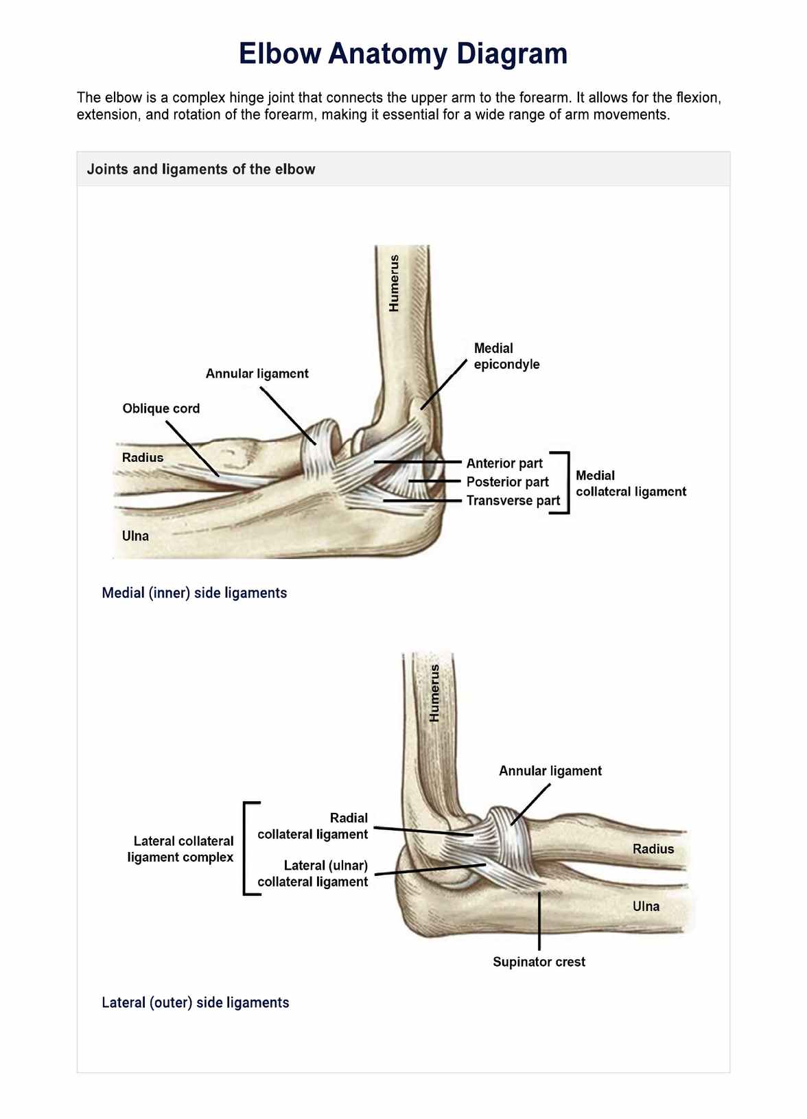
- Medial collateral ligament: A ligament that stabilizes the medial side (the inner side, facing the chest) of the elbow. This group is composed of three parts:
- Anterior part: Connects the medial epicondyle to the coronoid process.
- Posterior part: Connects the medial epicondyle to the olecranon.
- Transverse part: Connects the anterior and posterior parts of the medial collateral ligament.
- Annular ligament: Encircles the head of the radius and holds it in place within the radial notch of the ulna.
- Oblique cord: A small ligament that runs from the ulna to the radius just below the radial tuberosity.
- Lateral collateral ligament complex: A group of ligaments that stabilize the lateral side (the outer side) of the elbow. It comprises these two parts:
- Radial collateral ligament: Connects the lateral epicondyle to the annular ligament and radial notch.
- Lateral (ulnar) collateral ligament: Connects the lateral epicondyle to the ulna.
Bones
The elbow includes three major bones: the humerus, the radius, and the ulna.
Humerus
The humerus is the long bone of the upper arm, extending from the shoulder to the elbow.
- Lateral supracondylar ridge: A bony ridge on the lateral side of the humerus.
- Medial supracondylar ridge: A bony ridge on the medial side of the humerus.
- Radial fossa: A shallow depression on the anterior humerus that accommodates the radial head during flexion.
- Coronoid fossa: A depression on the anterior humerus that receives the coronoid process of the ulna during flexion.
- Lateral epicondyle: A bony prominence on the lateral side of the humerus.
- Medial epicondyle: A bony prominence on the medial side of the humerus.
- Capitellum/capitulum: A rounded eminence on the distal humerus that articulates with the head of the radius.
- Trochlea: A spool-shaped part of the humerus that articulates with the ulna.
- Lateral border: The outer edge of the humerus.
- Olecranon fossa: A large depression on the posterior humerus that accommodates the olecranon of the ulna during extension.
- Ulnar groove: A groove on the posterior aspect of the medial epicondyle through which the ulnar nerve passes.
Radius
The radius is one of the two long bones in the forearm, located on the lateral side (thumb side).
- Head of radius: The proximal end that articulates with the capitulum of the humerus and the radial notch of the ulna.
- Neck of radius: The narrow part just below the head of the radius.
- Radial tuberosity: A bony prominence below the neck where the biceps tendon attaches.
Ulna
The ulna is the longer of the two bones in the forearm, located on the medial side (pinky side).
- Ulnar tuberosity: A roughened area below the coronoid process where the brachialis muscle attaches.
- Coronoid process: A triangular projection on the anterior ulna that fits into the coronoid fossa of the humerus.
- Olecranon: The prominent bony projection of the ulna at the elbow.
- Supinator crest: A ridge on the lateral surface of the ulna where the supinator muscle attaches.