


What is the cervical spine?
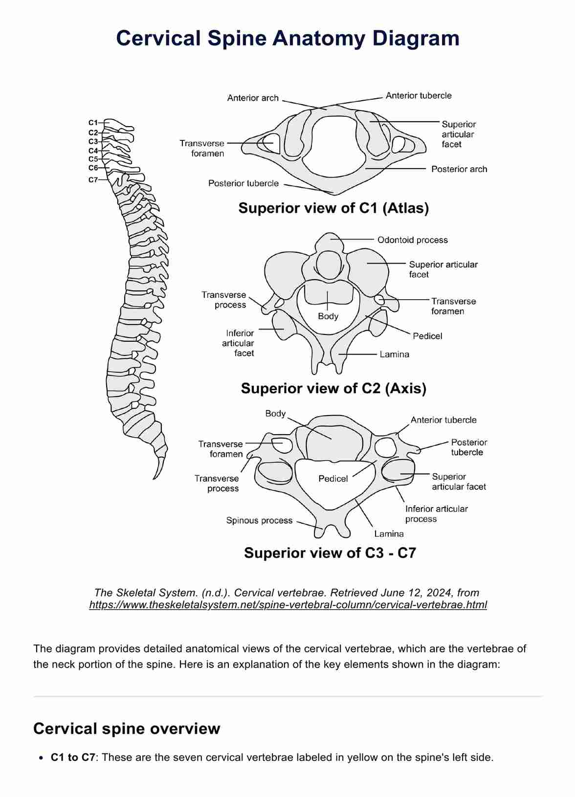
The cervical spine is the uppermost portion of the vertebral column, consisting of seven cervical vertebrae (C1-C7). Its main functions are to support the weight of the head, enable head and neck movement, and protect the spinal cord and vertebral arteries that pass through it.
The first two cervical vertebrae, the atlas (C1) and axis (C2), are specialized to allow for extensive rotation of the head. The remaining vertebrae (C3-C7) follow a more typical structure, with vertebral bodies separated by intervertebral discs that act as shock absorbers. Spinal nerves exit between each vertebral level.
The cervical spine also includes critical structures such as the anterior longitudinal ligament and posterior longitudinal ligament, which provide stability. The vertebral foramen is the passageway for the spinal cord, and injuries to this area can lead to significant spinal cord injury and chronic neck pain. Understanding the anatomy of the cervical spine is crucial for procedures like cervical spine surgery and for diagnosing conditions related to the cervical vertebrae and cervical spine ligaments.


