Axillary Nerve Block Anatomy Diagram

Our anatomy diagram enhances your understanding of the axillary nerve block. Download our free PDF template to guide your upper limb procedures.

By Olivia Sayson on Jul 15, 2024.

tick

Fact Checked by Ericka Pingol.

Use Template
Axillary Nerve Block Anatomy Diagram PDF Example
ToolbarShare uiAI Icon

What is the nerve axillary block?

The axillary nerve block is a crucial procedure in regional anesthesia, particularly for upper limb surgery involving the elbow, forearm, and hand. This axillary block targets the terminal branches of the brachial plexus as they pass through the axilla, providing adequate analgesia and anesthesia.

An ultrasound-guided axillary brachial plexus block allows direct visualization of the nerves, axillary artery, and needle, improving block success and safety compared to traditional landmark or nerve stimulation techniques. Understanding the axillary region's detailed anatomy is essential for effectively performing this ultrasound-guided block.

Axillary Nerve Block Anatomy Diagram Template

Download PDF Template

Axillary Nerve Block Anatomy Diagram Example

Download Example PDF

How does our Axillary Nerve Block Anatomy Diagram work?

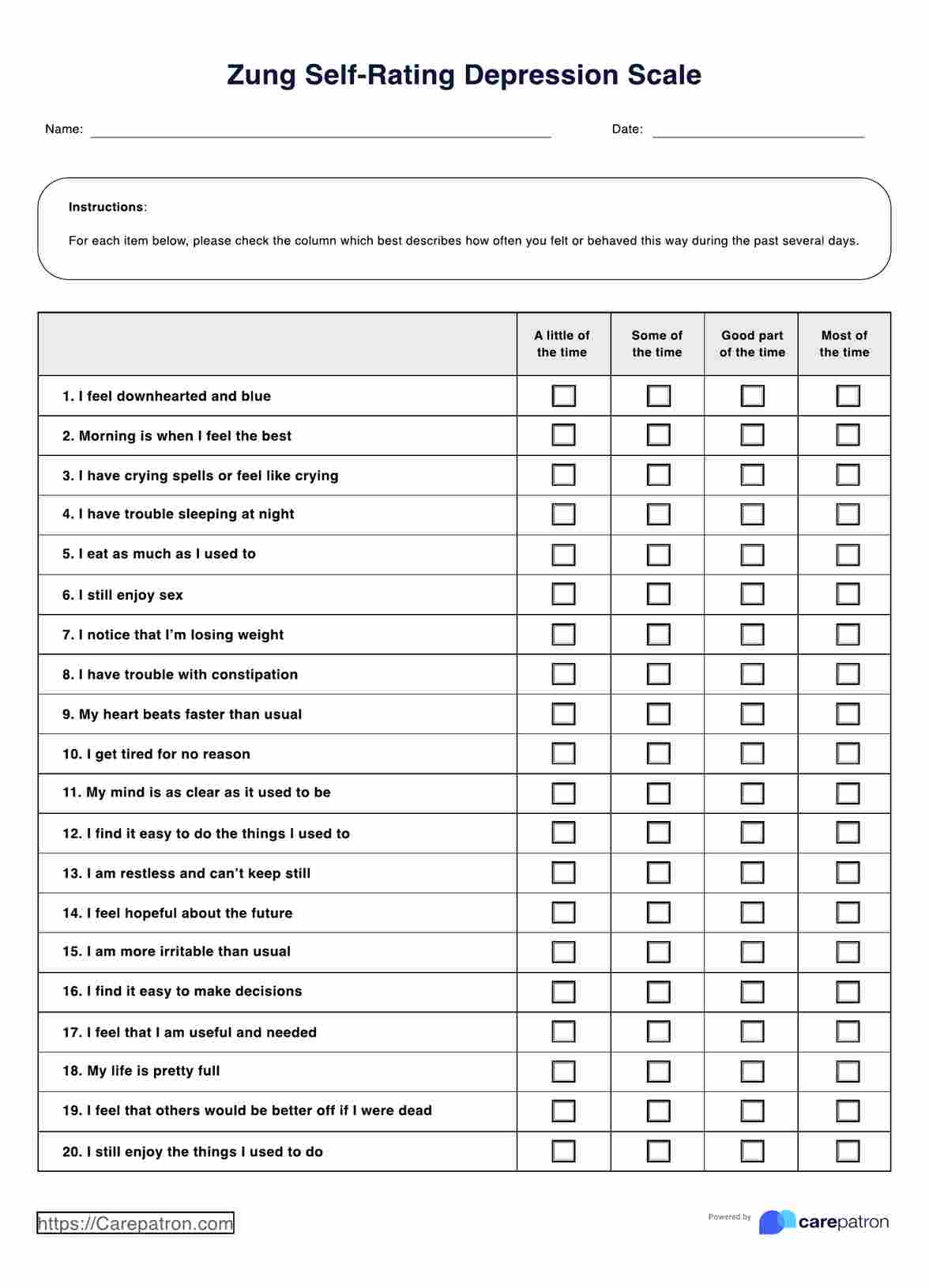
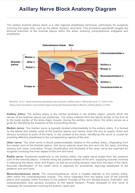
The Axillary Nerve Block Anatomy Diagram is a critical tool for understanding the precise locations of nerves and structures within the axillary region, essential for performing the procedure accurately. Our Axillary Nerve Block Anatomy Diagram provides a clear overview of the relevant structures for an ultrasound-guided axillary brachial plexus block, including:

  • Axillary artery
  • Median nerve (typically located superior and lateral to artery)
  • Ulnar nerve (inferior and medial to artery)
  • Radial nerve (posterior to artery)
  • Medial cord of brachial plexus

The diagram shows the typical anatomical arrangement and includes explanations of the parts of the anatomy for better understanding.

How will our diagram benefit healthcare professionals?

Our diagram is a valuable resource for anesthesiologists, surgeons, and medical trainees involved in upper limb surgeries. It is beneficial in:

  • Pre-procedural Planning: Aiding in the visualization of anatomical landmarks.
  • Educational Tool: Assisting in the teaching and training medical students and residents.
  • Clinical Practice: Enhancing the accuracy and safety of performing the axillary nerve block.
Why is understanding the anatomy essential for an axillary nerve block?

Commonly asked questions

Why is understanding the anatomy essential for an axillary nerve block?

Understanding anatomy is crucial because it helps practitioners accurately locate the nerves and surrounding structures, such as the axillary artery. This minimizes the risk of complications and increases the success rate of the block.
What structures are typically included in an Axillary Nerve Block Anatomy Diagram?

An Axillary Nerve Block Anatomy Diagram typically includes the axillary artery, median, and ulnar nerves, radial, and musculocutaneous nerve.

How can I use the Axillary Nerve Block Anatomy Diagram in clinical practice?

The diagram can be used as a visual aid during pre-procedural planning, helping to identify and understand the location of nerves and other critical structures.

Join 10,000+ teams using Carepatron to be more productive

One app for all your healthcare work