Understanding AC joint pathology

The acromioclavicular joint, or AC joint, is located at the top of the shoulder where the acromion (part of the scapula) meets the clavicle (collarbone). It allows for movement and stability in the shoulder region. However, like any other joint in our body, it can be affected by various pathologies.

One of the most common joint pathologies of the AC joint is osteoarthritis. Osteoarthritis is a degenerative disease that affects the cartilage in our joints, causing it to wear down over time. This can lead to pain, stiffness, and limited range of motion in the shoulder. In the case of AC joint osteoarthritis, the cartilage between the acromion and clavicle may wear down, causing the bone to rub against bone and resulting in pain and inflammation.

Other joint pathologies that can affect the AC joint include rheumatoid arthritis, which is an autoimmune disorder that causes inflammation in the joints, as well as injuries such as dislocations or fractures. These can disrupt the normal functioning of the joint and lead to pain and instability.

Printable AC Shear Test

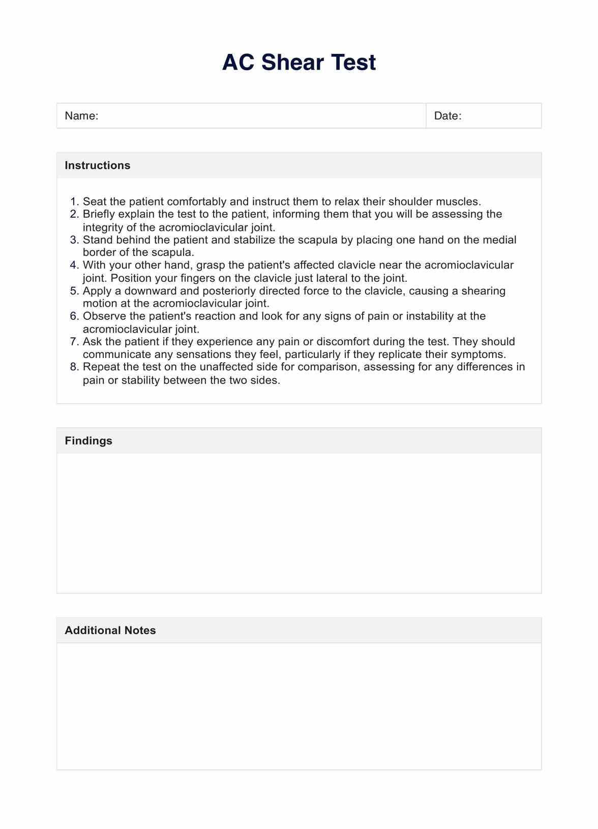
Download this AC Shear Test to assess the integrity of the AC joint and its surrounding structures.

Common symptoms of AC joint pathology

AC joint pathology can present itself in various forms, depending on the specific condition affecting the joint. However, some common symptoms may indicate an issue with the AC joint:

  • Pain or discomfort at the top of the shoulder
  • Difficulty lifting or moving the arm above the head
  • Swelling and tenderness around the AC joint
  • Limited range of motion in the shoulder
  • Clicking or grinding sensation in the joint
  • Weakness in the shoulder muscles

Individuals with AC joint pathology may also experience pain and discomfort while performing certain activities, such as reaching for objects or participating in sports that involve overhead movements.

What is the AC Shear Test?

The acromioclavicular shear test or AC Shear Test is a diagnostic test used to assess the integrity of the AC joint and its surrounding structures. It involves applying downward pressure on the acromion while simultaneously moving the arm in different positions. This test helps determine if there is any instability or damage to the AC joint, such as with an injury or pathology.

To perform the AC Shear Test, the patient is seated or standing with their arms relaxed at their sides. The physician stands behind the patient and places one hand on top of the acromion and the other hand on the elbow. Then, they apply downward pressure on the acromion while moving the arm in different directions, such as across the body or above the head.

If the patient experiences pain or movement in the AC joint during this test, it may indicate a problem with the joint, such as instability or damage to the ligaments. However, further imaging and diagnostic tests are usually needed to confirm a diagnosis.

AC Shear Test example (sample)

Our team has designed a user-friendly template to make it easy for you to perform the AC Shear Test. The template consists of easy-to-follow instructions and sections to jot down your findings and notes.

We also created a sample completed template, illustrating how you can use the template in your practice. Feel free to view the sample here or download a PDF copy for your reference.

Download this free AC Shear Test example here

AC Shear Test example (sample)

Results and interpretation

The results of the AC Shear Test can be interpreted based on the patient's response to the test. If the patient experiences pain or discomfort during the test, it may indicate a problem with the AC joint. The severity and location of pain can also provide valuable information about the specific issue.

For example, if there is pain on top of the acromion when pressure is applied, it may indicate damage to the AC ligament. If there is pain in the front of the shoulder or in the clavicle area, it may suggest instability or damage to other surrounding structures.

In some cases, a positive test may require further imaging tests, such as X-rays or MRI, to confirm a diagnosis and determine the extent of the injury or pathology.

The AC Shear Test is a simple, quick, and non-invasive test that can provide valuable information about the AC joint. It can be performed in a clinical setting with minimal equipment and does not require any special preparation from the patient.

However, this test may not always provide conclusive results. Factors such as patient positioning, muscle tension, and pain tolerance can affect its accuracy. Additionally, it is important to interpret the results in conjunction with other diagnostic tools, physical tests, and clinical findings.

Can the AC Shear Test produce both false positives and false negatives?

Commonly asked questions

Can the AC Shear Test produce both false positives and false negatives?

Yes, the AC Shear Test, like any diagnostic procedure, has a certain margin for error, and it is possible to encounter both false positives and false negatives. Factors that influence these discrepancies include the examiner's technique, the patient's response to pain, and the inherent limitations of the test itself. Supplementing this test with additional diagnostic tools and clinical assessments is crucial to ensure an accurate diagnosis.
How effective is the AC Shear Test in diagnosing isolated chronic acromioclavicular lesions?

The AC Shear Test can be particularly valuable for diagnosing isolated chronic acromioclavicular lesions, as it directly assesses the integrity of the AC joint by applying pressure to isolate this specific joint. However, the diagnostic value of this test increases when used alongside other evaluations since it can sometimes miss subtle chronic conditions or when the pathology involves surrounding structures.

What is the diagnostic value of the AC Shear Test?

The diagnostic value of the AC Shear Test lies in its ability to quickly screen for AC joint pathology through a simple and non-invasive procedure. While it is an effective initial assessment tool, its definitive diagnostic value is best achieved when combined with a comprehensive clinical examination, patient history, and potentially further imaging studies to confirm the extent and specifics of the injury or lesion.

Join 10,000+ teams using Carepatron to be more productive

One app for all your healthcare work