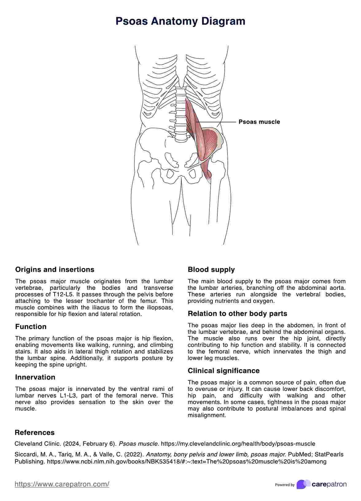
The psoas muscle plays a crucial role in various bodily movements, particularly in hip flexion and stabilization of the lumbar spine. Originating from the transverse processes of the lumbar vertebrae and extending to the anterior superior iliac spine, the psoas muscle, when combined with the iliacus muscle, forms the iliopsoas tendon.
Psoas Anatomy Diagram
Get access to a free Psoas Anatomy Diagram. Download the PDF and use as a handy reference for your practice.

Use Template
Psoas Anatomy Diagram Template
Commonly asked questions
The psoas muscle is located deep within the posterior abdominal wall, lying adjacent to the vertebral column. It traverses the bony pelvis and passes beneath the inguinal ligament before attaching to the lesser trochanter of the femur. The muscle's unique positioning allows it to interact with other important structures, such as the psoas minor muscle, quadratus lumborum muscle, and the anterior rami of the lumbar plexus.
Several conditions can impact the psoas muscle, including psoas syndrome, tendonitis, and muscle strain. These issues typically result from overuse, improper posture, or trauma in the region. Symptoms often manifest as lower back pain and difficulties in performing hip flexion, among others.


EHR and practice management software
Get started for free
*No credit card required
Free
$0/usd
Unlimited clients
Telehealth
1GB of storage
Client portal text
Automated billing and online payments